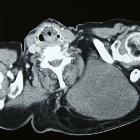
atypischer lipomatöser Tumor

Imaging
spectrum of abnormal subcutaneous and visceral fat distribution. Atypical lipomatous tumor in a 62-year-old male with a painless, firm, and mobile mass gradually increasing in the right back. a Coronal CT image shows a fat-containing inhomogeneous density mass (arrow). b On post-contrast fat-saturated T1-weighted image, the non-fatty lesion shows moderate enhancement (arrowheads). Surgically diagnosed as ALT with abundant fat necrosis

Differentiating
atypical lipomatous tumors from lipomas with magnetic resonance imaging: a comparison with MDM2 gene amplification status. Intramuscular lipomatous tumor in a 45-year-old female patient in the left gluteus muscles. (a) The axial T1 weighted image with fat saturation (FS) and (b) the axial T2 weighted image show a well-defined lipomatous tumor which was classified as a lipoma after resection. Another 65-year-old male patient showed an intramuscular lipomatous tumor reaching into the subcutaneous region of the left shoulder. (c) An axial T2 weighted and a (d) coronal T2 weighted image demonstrating pseudo-infiltrative margins of the tumor, which was classified as an ALT after resection

Differentiating
atypical lipomatous tumors from lipomas with magnetic resonance imaging: a comparison with MDM2 gene amplification status. A 41-year-old male patient with a lipomatous tumor (ALT) at the medial left sided thigh showing solid, non-lipomatous components within the tumor on a (a) coronal T1 weighted image with (b) contrast enhancement (> 1/3 of the tumor volume) on a coronal T1 weighted FS image. On the other hand, there is a 45-year-old female patient showing a lipomatous tumor (lipoma) on a (c) sagittal T2 weighted image without (d) contrast enhancement on the coronal T1 weighted FS image in the gluteus region on the left side

Differentiating
atypical lipomatous tumors from lipomas with magnetic resonance imaging: a comparison with MDM2 gene amplification status. Axial T2 weighted images of lipomatous tumors (a) without septa; (b) with thin septa (< 2 mm) and (c) with thick septa (> 2 mm)
 Assoziationen und Differentialdiagnosen zu atypischer lipomatöser Tumor:


