abnormale subkutane und viszerale Fettverteilung

Imaging
spectrum of abnormal subcutaneous and visceral fat distribution. A 23-year-old woman with congenital generalized lipodystrophy and fifth finger pain. Lytic lesion (arrowheads) was evident on (a) radiograph and (b) CT in the fifth middle phalange. c CT showed almost complete loss of fat tissue other than the palmar fat pad (arrow). MRI showed complete lack of bone marrow adipose tissue. Coronal (d) T2-weighted and (e) STIR images show almost the same signal. f T1-weighted image shows no adipose high signal intensity. An asterisk denotes the referred lytic lesion. g DIXON-based fat image shows almost complete loss of fat high signal in the bone marrow and soft tissue

Imaging
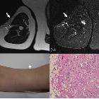
spectrum of abnormal subcutaneous and visceral fat distribution. A 59-year-old woman with post blunt trauma lipoatrophy on the right upper arm. a Axial T2-weighted and (b) STIR images show thinning of subcutaneous fat on the lateral aspects (arrows) with multiple high signal nodules (small arrows) showing small peripheral fat signal areas. c Photograph shows thinning of the lateral aspect of the upper arm with small hump (arrow). d Histologically fat necrosis with lipogranuloma was proven. Variably sized lipid vacuoles are surrounded by foam cells, foreign body-type (arrows), and Touton giant cells (arrowhead) in the resected lipogranuloma. A adipocytes, F fibrosis (hematoxylin-eosin stain, × 100)

Imaging
spectrum of abnormal subcutaneous and visceral fat distribution. Lipomatosis of the median nerve in an 11-year-old girl without symptoms. a Axial T2- and (b) coronal T1-weighted images reveal soft-tissue hypertrophy with predominance of fat in the radial side of the middle finger along with the neurovascular structure (arrows)

Imaging
spectrum of abnormal subcutaneous and visceral fat distribution. Spindle cell lipoma in a 68-year-old man with a painless mass on the posterior neck. a Axial T1-weighted image and b sagittal STIR image show a subcutaneous, encapsulated fatty mass (arrows) with amorphous non-fatty signal area (arrowheads) representing intermingled components such as collagen fibers, myxoid matrix, and vascular elements

Imaging
spectrum of abnormal subcutaneous and visceral fat distribution. Angiolipoma in a 29-year-old man on the left upper arm. a T1-weighted image shows a subcutaneous tiny mass with inhomogeneous high to intermediate signal intensity (arrow). b Fat-saturated T2-weighted image shows hyperintense signal with focal fat suppression (arrowhead) in the mass with connection to dilated subcutaneous vein (small arrow)

Imaging
spectrum of abnormal subcutaneous and visceral fat distribution. Superficial lipoma in a 60-year-old woman on the right shoulder. a T1-weighted image and (b) fat-suppressed T2-weighted show a homogeneous fatty mass (arrows) with a similar signal intensity to that of the adjacent subcutaneous fat but with a thin capsule and thin internal septa

Imaging
spectrum of abnormal subcutaneous and visceral fat distribution. A 47-year-old woman with semicircular lipoatrophy. a, b Linear horizontal depression on the lateral aspect of left thigh at 72 cm above the ground (arrows). She had a history of working leaning against the edge of her desk daily. c, d Coronal localizer image and STIR show loss of localized subcutaneous fat tissue with slight edematous change (circle)

Imaging
spectrum of abnormal subcutaneous and visceral fat distribution. A 60-year-old man with post-traumatic pseudolipoma on the right posterior neck. He had a history of carrying a mikoshi (Japanese portable shrine) daily. a Axial T1-weighted and b sagittal post-contrast fat-sat T1-weighted images show non-capsulated subcutaneous fat tissue proliferation on the right posterior neck (arrows). c Photograph of a mikoshi

Imaging
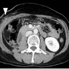
spectrum of abnormal subcutaneous and visceral fat distribution. A 59-year-old woman with post-surgery lipoatrophy. a CT image before operation shows calcified soft tissue density mass anterior to the right kidney (arrow). Right nephrectomy was done, and the lesion was pathologically diagnosed as dedifferentiated liposarcoma. b CT image one month after surgery shows fluffy opacity in the subcutaneous fat around the operated area (arrowhead) with abdominal wall muscle swelling. c CT image after 8 years shows local lipoatrophy (double arrow) with muscle atrophy

Imaging
spectrum of abnormal subcutaneous and visceral fat distribution. A 29-year-old man with Parry-Romberg syndrome. a Axial CT images show asymmetrically decreased subcutaneous fat on the left side (arrows). b Hypoplasty of the left orbit is also evident. c Volume-rendered three-dimensional reformation image of facial bones highlights bony asymmetry of the face. There is a perceivable asymmetry of the maxillary bone as well. d Coronal FLAIR image shows subcortical high signal intensity area (arrowhead)

Imaging
spectrum of abnormal subcutaneous and visceral fat distribution. Whole body MRI taken from the same CGL patient 10 years earlier. T1-weighted axial images at the level of (a) orbita, (b) axilla, (c) renal sinus, (d) pelvis, (e) thigh, and (f) sagittal image of foot show nearly complete absence of adipose tissue, which is however slightly preserved in the orbits, axillae, and soles (arrows)

Imaging
spectrum of abnormal subcutaneous and visceral fat distribution. A 48-year-old woman with deep morphea on her right thigh. a Axial fat saturated T2-weighted image shows hypersignal intensity in full thickness of subcutaneous fat on the anterior aspect (arrow). Increased signal intensity in gastrocnemius muscle (asterisks), muscle fasciae, and diffuse subcutaneous septal thickening are also demonstrated. b T1-weighted image clearly shows thinning of subcutaneous fat with cord-like low signal intensity (small arrows)

Imaging
spectrum of abnormal subcutaneous and visceral fat distribution. A 52-year-old man with Cushing disease. a Lateral skull radiography shows “buffalo hump” on the posterior neck (asterisk). Note the enlarged sella due to pituitary macroadenoma (arrowhead). b Frontal skull radiograph shows a rounded shape of the face, “moon face” (double arrows). c CT scanogram shows characteristic central obesity. d CT image at the level of bilateral renal hilum shows marked visceral fat deposition, in contrast to subcutaneous fat thinning (boxed arrows)

Imaging
spectrum of abnormal subcutaneous and visceral fat distribution. A 45-year-old man with multiple symmetrical lipomatosis with sleep apnea. a Axial T2-weighted and (b) T1-weighted image show abnormal subcutaneous fat deposition in the anterior (arrows) and posterior neck (asterisks). Note the increase of adipose tissue also observed in the posterior pharyngeal space (small arrows). c Post liposuction axial and (d) sagittal reconstructed CT 2 years later. Subcutaneous fat volume in the anterior neck is markedly decreased (arrows), whereas in contrast, the fat deposition in the posterior neck has worsened (asterisk)

Imaging
spectrum of abnormal subcutaneous and visceral fat distribution. A 66-year-old man with type I diabetes and insulin injection lipohypertrophy. a CT image at the level of belly button shows bilateral localized subcutaneous fat deposition on the anterior abdominal wall (arrows). b Volume-rendered three-dimensional reformation image show bilateral bulging of abdominal wall reflecting the insulin lipohypertrophy

Imaging
spectrum of abnormal subcutaneous and visceral fat distribution. A 47-year-old man with HALS. a Axial T2-weighted and (b) sagittal T1-weighted images show subcutaneous lipohypertrophy on the posterior neck (arrows). Marked loss of subcutaneous fat including buccal fat pad is shown in the face (arrowheads). c Volume-rendered three-dimensional reformation image shows apparent concave cheeks and periorbital hollowing

Imaging
spectrum of abnormal subcutaneous and visceral fat distribution. A 37-year-old man with binge eating disorder and overeating habitus (BMI = 53.2). a Chest radiograph shows bilateral pseudogynecomastia (black arrows). b CT of the chest and (c) abdomen shows increase of subcutaneous and visceral adipose tissue. Note the absence of extensive growth of mammary gland but pure fat deposition on the breast (white arrows)

Imaging
spectrum of abnormal subcutaneous and visceral fat distribution. A 48-year-old woman with serous atrophy of bone marrow from bulimia nervosa. a Coronal T1-weighted image demonstrates abnormal diffuse hypointense marrow signal with fracture of left femoral neck. b Coronal STIR demonstrates abnormal hyperintense signal in the bone marrow and surrounding subcutaneous or intermuscular tissue (arrows). c CT at the level of the femoral fracture (double arrow) shows depleted high attenuated fat tissue. d Histopathologically shrinkage of fat cells (black arrows), decrease of hematopoietic cells, and deposition of abundant amorphous extracellular gelatinous substances (asterisk) were observed (hematoxylin-eosin stain, × 100)

Imaging
spectrum of abnormal subcutaneous and visceral fat distribution. A 68-year-old man with pancreatic cancer. Initial CT in our hospital (a, b) shows massive low-density mass in the head of the pancreas (arrow). CT performed 14 months later (2 months before death) (c, d) shows depletion and increased attenuation of subcutaneous and visceral adipose tissue. Hepatic metastases (arrowheads) and copious ascites (asterisks) are also observed

Imaging
spectrum of abnormal subcutaneous and visceral fat distribution. Atypical lipomatous tumor in a 62-year-old male with a painless, firm, and mobile mass gradually increasing in the right back. a Coronal CT image shows a fat-containing inhomogeneous density mass (arrow). b On post-contrast fat-saturated T1-weighted image, the non-fatty lesion shows moderate enhancement (arrowheads). Surgically diagnosed as ALT with abundant fat necrosis
abnormale subkutane und viszerale Fettverteilung
Siehe auch:
- Adipositas
- Morel Lavallée Läsion
- Lipomatose
- atypischer lipomatöser Tumor
- Anasarka
- Anorexia nervosa
- subkutanes Lipom
- zirkumskripte Sklerodermie
- fibrolipomatöses Hamartom des Nervens
- progressive faziale Hemiatrophie
- Cushing-Syndrom
- Lipohypertrophie unter Insulintherapie
- Angiolipom
- Lipodystrophie
- Lipoatrophie
- Spindelzell-Lipom
- HIV-1 highly active antiretroviral therapy (HAART)-associated lipodystrophy syndrome (HALS)

 Assoziationen und Differentialdiagnosen zu abnormale subkutane und viszerale Fettverteilung:
HIV-1 highly
active antiretroviral therapy (HAART)-associated lipodystrophy syndrome (HALS)


