Nasal bone hypoplasia
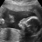
Hypoplasia of the nasal bone refers to a sonographic observation where the fetal nasal bone appears smaller by varying degrees.
There is a spectrum of nasal bone hypoplasia, at one end of which is the relatively easily identified absent nasal bone. The other end of the spectrum is considerably harder to define, particularly when used as a screening tool (see below).
Epidemiology
0.5-1.2% of normal fetuses have been found to have a hypoplastic nasal bone on a routine 2 trimester scan, compared to 43-62% of fetuses with Down syndrome .
Associations
- Down syndrome: nasal bone hypoplasia has emerged as one of the strongest morphological markers of trisomy 21 to date
- it relates to the phenotypical observation that individuals with Down syndrome have short noses; as well as a growing body of supportive radiological data
- fetal warfarin syndrome is a rare association
Radiographic assessment
Antenatal ultrasound
The nasal bone is best assessed in the second trimester, and its measurement is a standard component of a routine 2 trimester ultrasound. It is assessed on a midsagittal view of the fetal face. Ideally, three echogenic lines should be seen.
The difficulty in defining nasal bone hypoplasia has historically lead to the development of various criteria, based on measurements such as BPD: nasal bone ratio , gestational age-adjusted nasal bone length, or a single cut-off definition (2.5 mm) .
Many practices now favor the use of data sets that define normal nasal bone length by gestational age, such as that by Obido et al. published in 2007 . More recently Mogra et al. published a normative data set based on an Australian multiethnic population, and this has now superseded the earlier data sets in some Australian centers .
See also
- soft markers on antenatal ultrasound
- antenatal features of Down syndrome
- automatic online fetal nasal bone length calculator from www.perinatology.com
Siehe auch:
- Down-Syndrom
- absent nasal bone
- antenatal features of Down syndrome
- Fetales Warfarinsyndrom
- soft markers on antenatal ultrasound
und weiter:

 Assoziationen und Differentialdiagnosen zu hypoplastic nasal bone:

