absent nasal bone


In a fetal sonographic assessment, an absent nasal bone is a feature that can sometimes be used as a surrogate marker for fetal aneuploidy.
Radiographic assessment
Antenatal ultrasound
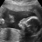
It is assessed on a midline sagittal view. In this section, the nasal bone is often seen as a bright echogenic line. It is best visualized at around 11 to 14 weeks of gestation (1trimester). A magnified image may assist in visualization.
When the mid-sagittal view is difficult to assess, some authors suggest a coronal view of the fetal face to look for paired echogenic structures located at the upper tip of the retronasal triangle .
Significance
When the nasal bone is absent at 11 to 12 weeks, while the other ultrasound markers and serum biochemistry are normal; a follow-up scan after a week is suggested.
The incidence of an absent nasal bone is related to nuchal translucency (NT), crown-rump length (CRL), and ethnic origin, as well as aneuploidy. It is more common with increased NT, smaller CRL measurements, and in fetuses of Afro-Caribbean parents.
The reported prevalence range of an absent nasal bone on ultrasound for euploid as well as the following aneuploidy states are
- euploid: nasal bone absent in 0.5-3%
- trisomy 21: nasal bone absent in 60-73%
- trisomy 18: nasal bone absent in 53-57%
- trisomy 13: nasal bone absent in 32-45%
- Turner syndrome: nasal bone absent in 9%
See also
Siehe auch:
und weiter:


 Assoziationen und Differentialdiagnosen zu absent nasal bone:


