Cardiac valves
Cardiac
valves • Cardiomediastinal anatomy on chest radiography (annotated images) - Ganzer Fall bei Radiopaedia


desc}} ==
{{Information |description = |Other fields 1 = {{Information field|Name={{Ucfirst:{{Plate}}}}|Value=495}} |date = {{Other date|before|1858}} |source = *{{cite book |year=1918 |author=[[:en:Henry Gray|Henry Gray]] |title=Anatomy of the Human Body }} (See "{{Section header|Book}}" section below) *{{Gray"s Anatomy/link|495}} |author = {{Creator:Henry Vandyke Carter}} |other_versions = |permission = }} == {{Section header|Book}} == {{Gray"s Anatomy}} =={{int:license-header}}== {{PD-scan|PD-old-100-1923}} [[Category:Gray"s Anatomy plates|0495]] [[Category:He

desc}} ==
{{Information |description = |Other fields 1 = {{Information field|Name={{Ucfirst:{{Plate}}}}|Value=1218}} |date = {{Other date|before|1858}} |source = *{{cite book |year=1918 |author=[[:en:Henry Gray|Henry Gray]] |title=Anatomy of the Human Body }} (See "{{Section header|Book}}" section below) *{{Gray"s Anatomy/link|1218}} |author = {{Creator:Henry Vandyke Carter}} |other_versions = |permission = }} == {{Section header|Book}} == {{Gray"s Anatomy}} =={{int:license-header}}== {{PD-scan|PD-old-100-1923}} [[Category:Gray"s Anatomy plates|1218]] [[Category:Heart (organ)]] [[Category:Sternum]] [[Category:Ribs

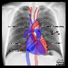
Cardiac
valves • Sectional cardiac anatomy (creative commons illustration) - Ganzer Fall bei Radiopaedia

Cardiac
valves • Mitral and aortic valve replacements - Ganzer Fall bei Radiopaedia

Cardiac
valves • Cardiac valves (illustration) - Ganzer Fall bei Radiopaedia

Cardiac
valves • Mitral valve replacement - Ganzer Fall bei Radiopaedia

Cardiac
valves • Cardiac fibrous skeleton (Gray's illustration) - Ganzer Fall bei Radiopaedia
The four cardiac valves direct the flow of blood through the heart during the cardiac cycle.
Gross anatomy
The heart valves are located in the cardiac fibrous skeleton:
- two are atrioventricular (AV) valves: the right-sided tricuspid valve (TV) and left-sided mitral (bicuspid) valve (MV)
- open during diastole to direct blood flow from the atria to the ventricles
- close during systole to prevent regurgitation back into the atria from the ventricles
- are attached to papillary muscles via chordae tendineae
- two are semilunar valves: the right-sided pulmonary valve (PV) and left-sided aortic valve (AV)
- open during systole to direct blood flow from the contracting ventricles through the right ventricle and left ventricle outflow tracts to the pulmonary trunk and ascending aorta, respectively
- close during diastole to prevent regurgitation back into the ventricles from the pulmonary trunk and ascending aorta
- these valves do not have chordae tendineae or papillary muscles
It is best to list the four valves in order of the series that blood travels through the heart:
See also
Siehe auch:


 Assoziationen und Differentialdiagnosen zu Atrioventricular valves (AVs):
