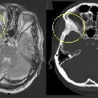
Fibröse Dysplasie im Clivus

Ausgedehnte
kraniofaziale fibröse Dysplasie, links partiell entlastet mit Kalottenersatz.

Fibröse
Dysplasie im Clivus: Typisches milchglasartiges Verdichtungsmuster. Hier allenfalls minimale Expansion (kann bei FD deutlicher sein) ohne arrosives Muster. Der Befund ist nicht histologisch bestätigt, aber in seinem computertomographischen Muster typisch. Insgesamt ist die Lokalisation im Clivus bei der kraniofazialen Form der Fibrösen Dysplasie jedoch selten. Links eine sagittale Rekonstruktion aus einer CT-Angiographie, rechts eine native, axiale Computertomographie. Die Untersuchung erbrachte die Fibröse Dysplasie als zufälligen Nebenbefund.

Ausgedehnte
kranialfaziale fibröse Dysplasie mit Beteiligung des Clivus und des linken Felsenbeins: Computertomografie und Magnetresonanztomografie (oben T2, unten T1). Typische milchglasartige Struktur der fibrös ersetzten Knochenanteile mit einigen lytischen Anteilen und Verbreiterung. In der Magnetresonanztomografie hypointense Darstellung des Felsenbeins sowohl in T1 wie auch in T2.

Fibrous
dysplasia • Fibrous dysplasia of skull base - Ganzer Fall bei Radiopaedia

Fibrous
dysplasia for radiologists: beyond ground glass bone matrix. CT and MRI in craniofacial fibrous dysplasia (FD). a CT demonstrates mixed sclerotic FD lesions involving the skull base (red arrows). b Lesions demonstrate intermediate signal intensity on T1 weighted MRI (green arrows). On T2 weighted images lesions demonstrate heterogeneous hypointense/intermediate signal intensity (blue arrows). d FD lesions show slightly heterogeneous enhancement (yellow arrows)

Fibrous
dysplasia for radiologists: beyond ground glass bone matrix. Optic nerves in craniofacial fibrous dysplasia (FD) in two different patients. a Extensive FD involving most of the facial bones and skull. Optic canals are narrowed but patent. b–d Expansile bone lesions in the left frontal bone, left sphenoid bones, ethmoid bone, and body of the sphenoid bone with marked narrowing and deformity of the left optic canal, causing left-sided blindness (red arrows). The right optic canal is narrowed (blue arrows). Vision in the right eye is preserved

Fibrous
dysplasia for radiologists: beyond ground glass bone matrix. Pituitary adenoma in craniofacial fibrous dysplasia (FD). a, b A patient with craniofacial FD (blue arrows) with pituitary macroadenoma (red arrows) on CT (a) and T1 contrast-enhanced MRI (b)

Fibrous
dysplasia for radiologists: beyond ground glass bone matrix. The location-based difference in appearance of fibrous dysplasia (FD) lesions. a Craniofacial FD demonstrates dense, sclerotic lesions (green arrow). b Lesions in the long bones and axial skeleton are typically lucent. There is a typical lesion in the proximal femur with characteristic lucent ground glass appearance and shepherd’s crook deformity (blue arrow). c Mixed radiolucent/lytic FD lesions in the skull

Craniofacial
fibrous dysplasia • Fibrous dysplasia - Ganzer Fall bei Radiopaedia

Fibrous
dysplasia • Fibrous dysplasia - base of skull - Ganzer Fall bei Radiopaedia

Craniofacial
fibrous dysplasia • Severe fibrous dysplasia of the skull base and face - Ganzer Fall bei Radiopaedia

Craniofacial
fibrous dysplasia • Fibrous dysplasia - Ganzer Fall bei Radiopaedia
Die isolierte Fibröse Dysplasie im Clivus ist eine eher seltene Lokalisation der kraniofazialen fibrösen Dysplasie, während eine Mitbeteiligung des Clivus bei ausgedehnteren Dysplasien der Schädelbasis häufiger vorkommt. Wenn die isolierte Form das typische Milchglasbild zeigt, wird sie in vielen Fällen dennoch als solche identifizierbar sein. Dabei ist die Computertomographie spezifischer als die MRT. Siehe hierzu den Hauptartikel zur Fibrösen Dysplasie.
Siehe auch:

 Assoziationen und Differentialdiagnosen zu Fibröse Dysplasie im Clivus:


