Abdominal cocoon


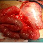


Encapsulating peritoneal sclerosis is a rare benign cause of acute or subacute small bowel obstruction. It is characterized by total or partial encasement of the small bowel within a thick fibrocollagenous membrane.
Terminology
The condition was originally termed abdominal cocoon. The condition is popularly known as sclerosing encapsulating peritonitis, however, this is somewhat of a misnomer as inflammation is not always present . It has also been known as sclerosing peritonitis, encapsulating peritonitis, and peritonitis chronica fibrosa incapsulata.
Epidemiology
Encapsulating peritoneal sclerosis can occur at any age, with reports ranging from 2-day neonate to 82 years .
Clinical presentation
The presentation is non-specific and patients may present with vomiting, abdominal pain and/or a subacute bowel obstruction .
Pathology
It can be idiopathic or secondary due to:
- continuous ambulatory peritoneal dialysis (prevalence ~0.7%)
- tuberculosis
- peritoneovenous or ventriculoperitoneal shunts
- treatment with practolol
Various abdominal disorders such as tuberculosis, sarcoidosis, familial Mediterranean fever, gastrointestinal malignancy, ovarian carcinoma , protein S deficiency, liver transplantation, fibrogenic foreign material, and luteinised ovarian thecomas are the other rare causes.
Radiographic features
Plain radiograph
Abdominal radiographic appearances are nonspecific and may be normal or may show:
- gas-fluid levels similar to those in patients with any other cause of small-bowel obstruction
- the wall of the "cocoon" may calcify
Ultrasound
- clumped bowel loops
- trilaminar appearance of a hyperechoic membrane, hypoechoic bowel wall and hyperechoic bowel contents
- ascites may be present
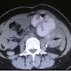
CT
In the appropriate clinical setting, recognition of the entire dilated small bowel at the center of the abdomen and encased within a thick fibrocollagenous membrane, as though it were in a cocoon, is diagnostic of sclerosing encapsulating peritonitis. The other imaging findings may include:
- enhancing peritoneum, thickened >2 mm
- signs of small intestinal obstruction
- fixation of intestinal loops
- ascites or localized fluid collections (especially interbowel)
- bowel wall thickening
- peritoneal or mural calcification
- calcified and/or reactive adenopathy
MRI
MRI will demonstrate the same features as CT, although it may better discriminate between thickened bowel and the peritoneal membrane than CT .
Differential diagnosis
Encapsulating peritoneal sclerosis may be confused with congenital peritoneal encapsulation, which is characterized by a thin accessory peritoneal sac surrounding the small bowel.
Siehe auch:
- ventrikuloperitonealer Shunt
- intraabdominelle Tuberkulose
- Peritonitis
- peritoneale Dialyse
- Peritonitis bei Peritonealdialyse
- zystische peritoneale Veränderungen
- practolol
und weiter:


 Assoziationen und Differentialdiagnosen zu Peritonitis chronica fibrosa incapsulata:


