Achilles tendon xanthoma

Multiple
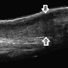
tendon xanthomas in a child with familial hypercholesterolaemia. Panoramic ultrasound image of right achilles tendon along its long axis (arrows). Diffuse thickening of achilles tendon is noted with loss of fibrillary architecture.
Multiple
tendon xanthomas in a child with familial hypercholesterolaemia. Panoramic ultrasound image of left Achilles tendon along its long axis (arrows). Diffuse thickening of achilles tendon is noted with loss of fibrillary architecture.
Achilles tendon xanthomata are painless soft tissue masses occurring most commonly at the distal portion of the tendon and are usually bilateral and symmetrical.
Pathology
Characterized by localized accumulation of lipid-laden macrophages, inflammatory cells and giant cells secondary to cholesterol deposition in tissue.
Causes
- familial hypercholesterolemia
- type 2 and 3 hypoproteinaemias
- cerebrotendinous xanthomatosis
Radiographic features
Plain radiograph
- thickening of the Achilles tendon
- no calcifications
- other tendon or periarticular soft tissue nodularity may be present
- often bilateral and symmetrical
Ultrasound
- an AP thickness of the tendon >7 mm in males and >6 mm in females
- tendon is more uniformly thickened as opposed to fusiform as seen in tendinopathy
- loss of normal tendon appearance with multiple hypoechoic foci within the tendon but not fluid dark
- often bilateral and symmetric
- other ankle flexor and extensor tendons can be involved
MRI
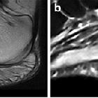
Shows increased AP diameter of the distal tendon which is often uniform, not fusiform. Loss of the normal anterior concavity of the tendon can also be seen.
Signal characteristics include.
- intermediate signal on T1- and T2-weighted sequences
- higher T1- and T2-weighted signal compared to the normal tendon
- T2 signal will not be 'fluid' bright as seen in partial-thickness tears
- striated appearance in sagittal sequences due to interposition of xanthoma between normal tendon fibers
- speckled appearance of the tendon in axial sequences
Other feautures
- often bilateral and symmetric
- other ankle flexor and extensor tendons can be involved
Differential diagnosis
Consider
- tophaceous gout
- chronic tendon degeneration (e.g. mucoid)
- tendinosis
- partial-thickness tears
- giant cell tumor of the tendon shealth
Siehe auch:


 Assoziationen und Differentialdiagnosen zu Xanthome der Achillessehne: