AIDS-related diffuse large B-cell lymphoma

Spectrum of
imaging findings in AIDS-related diffuse large B cell lymphoma. Chest radiograph of a 19-year-old HIV-positive patient with low CD4 count. He presented with cough and dyspnea and was initially being evaluated for possible atypical pneumonia. The chest radiograph showed multiple focal opacities predominantly in the lower lung zones. On CT, there were lung parenchymal nodules/masses of variable sizes which were also intensely FDG avid on PET CT. There was extensive lymphoma involvement elsewhere including in the liver, spleen (splenic activity higher than liver background), and multiple nodal sites above and below the diaphragm. Biopsy of the liver lesion was consistent with DLBCL

Spectrum of
imaging findings in AIDS-related diffuse large B cell lymphoma. Coronal reformats of a CECT abdomen and pelvis of a recently diagnosed HIV-positive patient who presented with abdominal pain, diarrhea, and weight loss show long-segment bowel thickening involving the distal ileum and caecum (long arrow). There was also mesenteric lymphadenopathy, with some necrotic nodes, (short slender arrow) and a moderate ascites (short fat arrow). There were also enlarged centrally necrotic right inguinal lymph nodes

Spectrum of
imaging findings in AIDS-related diffuse large B cell lymphoma. Axial CT images axial contrast-enhanced CT image and liver ultrasound of a patient with HIV-associated primary hepatic lymphoma. The contrast-enhanced CT shows a homogeneous soft tissue attenuation mass which does not show significant enhancement, and with fear preservation of the surrounding liver architecture. The mass displaces or encases the hepatic veins but does not invade vasculature or lead to tumor thrombus. On ultrasound, it appears as a fairly homogenous mass hypoechoic to the liver parenchyma. There is no increase vascularity within the mass or in its periphery

Spectrum of
imaging findings in AIDS-related diffuse large B cell lymphoma. Coronal and axial CECT images of the abdomen (a and b) show hepatomegaly and a heterogenous hypoattenuating mass infiltrating through most of the right lobe of the liver. The infiltrative liver mass had more focal hypoattenuating areas (fat arrow). Aortocaval lymph nodes are also noted (short arrow). Axial CT through the pelvis in bone window (c) shows a permeative process involving the left femoral head and neck and associated pathological fracture

Spectrum of
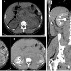
imaging findings in AIDS-related diffuse large B cell lymphoma. Ultrasound image of the left kidney in a HIV-positive male demonstrates heterogeneously echogenic areas in the medulla extending to the collecting system. Unenhanced CT KUB showed diffuse thickening of the renal pelvis with hyperdense soft tissue material relative to the renal parenchyma. Heterogenous infiltration of the kidney is seen on the postcontrast CT images in arterial and urographic phases

Spectrum of
imaging findings in AIDS-related diffuse large B cell lymphoma. CECT of the pelvis in soft tissue window (a) and bone window (b). A mixed lytic and sclerotic process involving the sacrum and extending to the iliac bones through the sacroiliac joints is seen. There are associated pathological fractures and a large extra osseous soft tissue component. There are also enlarged necrotic bilateral pelvic sidewall and inguinal lymph nodes

Spectrum of
imaging findings in AIDS-related diffuse large B cell lymphoma. A 46-year-old female patient who had pancreatic lymphoma (a) had a negative PET CT at the end of treatment (b and c). Nine months later, there was local disease recurrence at the pancreatic head on follow-up FDG PET CT scan (d and e)

Spectrum of
imaging findings in AIDS-related diffuse large B cell lymphoma. Newly diagnosed HIV-positive patient who had CD4 count of 151 presented with primary gastric lymphoma. He was treated with R-CHOP regimen complicated by severe neutropenic sepsis. Intrathecal chemotherapy was deferred to cycle 3 due to poor performance. Interim PET/CT showed good treatment response of the gastric tumor but there was refractory neuraxial disease seen as FDG avid conus medullaris

Spectrum of
imaging findings in AIDS-related diffuse large B cell lymphoma. This patient with extensive retroperitoneal and renal disease (a and b) showed excellent response to treatment in the interim PET CT (c, d, and e) after two cycles of R-CHOP. There was however early relapse with bulky disease developing in the retroperitoneum and the scrotum (f)

Spectrum of
imaging findings in AIDS-related diffuse large B cell lymphoma. Axial CT image (a) demonstrates bilateral axillary lymphadenopathy, proven to be DLBCL after biopsy. The patient successfully completed treatment but developed CNS symptoms 1 year later. MRI of the brain (b) showed a large heterogenous mass in the left cerebral hemisphere which biopsy was proven to be lymphoma. A previous MRI brain (c) was negative for CNS involvement
AIDS-related diffuse large B-cell lymphomas are one of the immunodeficiency-associated CNS lymphomas, and in western countries represented a dramatic increase in primary CNS lymphoma during the HIV/AIDS epidemic of the 1980s, although the incidence is likely lower in patients treated with HAART .
Pathology
Like other immunodeficiency-associated CNS lymphomas, AIDS-related lymphomas tend to be EBV-related .
Radiographic features
Imaging is similar to other immunodeficiency-associated CNS lymphomas, with tumors more likely to be necrotic, heterogeneous and multifocal .
Differential diagnosis
A particularly challenging differential diagnosis is cerebral toxoplasmosis. See cerebral toxoplasmosis vs primary CNS lymphoma.

