Arteriovenous fistulae


An arteriovenous fistula (AVF) is an abnormal connection between an adjacent artery and vein. Unlike an arteriovenous malformation (AVM), these are frequently acquired lesions, rather than developmental abnormalities.
Pathology
Arteriovenous fistulas have a number of etiologies. They can be iatrogenic in origin, particularly with percutaneous procedures, when a needle passes through both an artery and vein. They may also occur when an aneurysmal artery ruptures into an adjacent vein (as can happen with coronary artery aneurysms).
The most common arteriovenous fistula is intentional: surgically-created arteriovenous fistulas in the extremities are a useful means of access for long-term haemodialysis - See haemodialysis arteriovenous fistula.
Locations
These can occur in multiple locations with the more common ones having separate articles as below
- cerebral arteriovenous fistula
- scalp arteriovenous fistula (also called a cirsoid aneurysm)
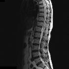
- spinal arteriovenous fistula
- coronary arteriovenous fistula
- renal arteriovenous fistula
Radiographic features
Ultrasound
- a fistula may be visualized directly, with an abnormal high-velocity connection between the artery and vein
- even if the fistula cannot be visualized directly, changes in the artery upstream from the fistula and the vein downstream from the fistula can establish a diagnosis:
- increased diastolic arterial flow due to its connection to the low-resistance vein
- arterialization of the vein downstream from the fistula (abnormal arterial pulsatility in the vein).
- enlargement of the downstream vein due to the increased volume of flow
- color bruit artifact in the adjacent soft tissues due to turbulent flow
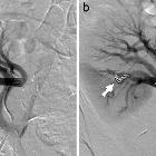
Angiography/DSA
- will show abnormal early filling of an adjacent vein in the region of the AVF
CTA/MRA
- abnormal early attenuation/intensity of a vein can prompt a search for a point of arteriovenous fistulization upstream. Time-resolved imaging sequences in MRI (e.g. TRICKS or TWIST) may be helpful.
Siehe auch:
- arteriovenöse Malformationen der Lunge
- spinale durale arteriovenöse Fistel
- arteriovenöse Fistel der Niere
- kraniale durale AV-Fistel
- Arteriovenöse Fisteln der Extremitäten
- iatrogenic arteriovenous fistula
- post-traumatic arteriovenous fistula
- arteriovenous fistula - treatment by embolisation
und weiter:


 Assoziationen und Differentialdiagnosen zu Arteriovenöse Fistel: