Bandverletzungen am Ellenbogen

The elbow:
review of anatomy and common collateral ligament complex pathology using MRI. Modified oblique coronal plane of the elbow used to optimally visualize the collateral ligaments. a Sagittal T1-weighted MRI showing the modified oblique coronal plane (multiple white lines) oriented 20–30° posterior to the long axis of the humeral diaphysis. b Coronal T1-weighted MRI showing the intact lateral ulnar collateral ligament (white asterisks)

The elbow:
review of anatomy and common collateral ligament complex pathology using MRI. Three different patients with lateral elbow pain. Coronal FS PD-weighted MRI (a–c) showing common extensor tendinosis (long arrow), common extensor tendon partial-thickness tear (arrowhead), and common extensor tendon full-thickness tear (short arrow)

The elbow:
review of anatomy and common collateral ligament complex pathology using MRI. A 25-year-old man with pain and medial instability after a fall on an outstretched hand. Axial T1-weighted MRI (a) showing the intact right posterior bundle of the medial collateral ligament complex of a healthy volunteer (white arrowhead). Axial FS PD-weighted MRI (b) showing an acute tear of the left posterior bundle of the medial collateral ligament complex (white arrow), inflammatory changes in the soft tissue of cubital tunnel (white asterisk), displacement and flattening of the ulnar nerve (yellow arrow), and joint effusion (black asterisks)

The elbow:
review of anatomy and common collateral ligament complex pathology using MRI. Posterolateral rotatory instability, stage 3B. Consecutive coronal FS T1-weighted direct MR arthrographic images (a–f) showing a proximal partial-thickness tear of the anterior bundle of the medial collateral ligament complex (short arrows), proximal complete tear of the lateral ulnar collateral (long white arrows), and partial-thickness tear of the radial collateral ligament proper (white arrowhead)

The elbow:
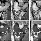
review of anatomy and common collateral ligament complex pathology using MRI. Posterolateral rotatory instability, stage 3B. Consecutive coronal T1-weighted MRI (a–c), and consecutive coronal FS PD-weighted MRI (d–f) showing an acute proximal full-thickness tear of the lateral ulnar collateral ligament and radial collateral ligament (white arrows) and anterior bundle of the medial collateral ligament complex sprain (white arrowheads)

The elbow:
review of anatomy and common collateral ligament complex pathology using MRI. Posterolateral rotatory instability, stage 2. Coronal FS PD-weighted MRI showing a complete detachment of the proximal lateral ulnar collateral ligament, the proximal radial collateral ligament proper, and the common extensor tendon (white short arrow), with retraction of the lateral ulnar collateral ligament (white arrowhead) and common extensor tendon (long white arrow). Note the extravasation of the joint fluid through the tear (black asterisk)

The elbow:
review of anatomy and common collateral ligament complex pathology using MRI. A 50-year-old man with elbow pain and decreased range of motion due to an apophyseal injury sustained before physeal closure. Axial T1-weighted MRI (a) and axial FS PD-weighted MRI (b) showing chronic non-union fracture of the medial epicondyle and heterotopic ossification along the anterior band of the medial collateral ligament (white arrows). Note stress reaction, manifest as bone marrow edema (white asterisk)

The elbow:
review of anatomy and common collateral ligament complex pathology using MRI. A 12-year-old left-handed baseball pitcher with medial epicondyle pain. Axial T1-weighted MRI (a), axial FS PD-weighted MRI (b), coronal FS PD-weighted MRI (c), and coronal T1-weighted MRI (d) showing a widening of the medial epicondylar physis (white arrows) and bone marrow edema (white asterisks)

The elbow:
review of anatomy and common collateral ligament complex pathology using MRI. Axial illustrations with superior view demonstrating the progression of the structures injured in the posterolateral rotatory instability

The elbow:
review of anatomy and common collateral ligament complex pathology using MRI. Anterior view of the elbow demonstrating the progression of the structures injured in a posterior dislocation, from lateral to medial. a Normal. b Proximal disruption of the lateral ulnar collateral ligament (white asterisk). c Proximal disruption of the lateral ulnar collateral ligament and the radial collateral ligament (black asterisk). d Anterior capsule tear (white arrow). Lateral ulnar collateral ligament (LUCL). Radial collateral ligament (RCL). Annular ligament (AL). Anterior bundle of the medial collateral ligament complex (A-MCL)

The elbow:
review of anatomy and common collateral ligament complex pathology using MRI. Posterolateral rotatory instability, stage 1. Coronal FS PD-weighted MRI (a, b) and coronal T1-weighted MRI (c, d) showing an avulsion of the proximal lateral ulnar collateral ligament and retraction of the osseous fragment (white arrows). Bone marrow edema is seen in the capitellum (white arrowheads) and radial head (white asterisks)

The elbow:
review of anatomy and common collateral ligament complex pathology using MRI. Normal appearance of reconstructed medial collateral ligament (docking technique). Consecutive coronal T1-weighted MRI (a–c) and coronal FS PD-weighted MRI (d). The intact graft (blue arrows) extends from humeral tunnel (blue arrowheads) to ulnar tunnel (yellow arrowheads)

The elbow:
review of anatomy and common collateral ligament complex pathology using MRI. Posterolateral rotatory instability, stage 3B. Consecutive coronal FS T1-weighted direct MR arthrographic images (a, b), sagittal FS T1-weighted direct MR arthrographic image (c), and consecutive axial FS T1-weighted direct MR arthrographic images (d, e) showing a proximal complete tear of the lateral ulnar collateral ligament and radial collateral ligament proper (white short arrows), diffuse partial-thickness tear of the anterior bundle of the medial collateral ligament complex (white arrowheads), and a non-displaced fracture of the coronoid process (long white arrows)

The elbow:
review of anatomy and common collateral ligament complex pathology using MRI. Posterolateral rotatory instability, stage 3C. Consecutive coronal FS T1-weighted direct MR arthrographic images (a–c), and consecutive axial FS T1-weighted direct MR arthrographic images (d–f) demonstrating a full-thickness tear of the anterior bundle of the medial collateral ligament complex (yellow arrow), proximal full-thickness tear of the common flexor tendon (short white arrows), proximal full-thickness tear of the common extensor tendon (white arrowheads), proximal complete tear of the lateral ulnar collateral ligament and radial collateral ligament proper (long white arrows), and a complete tear of the posterior bundle of the medial collateral ligament complex (yellow arrowheads)

The elbow:
review of anatomy and common collateral ligament complex pathology using MRI. Axial FS PD-weighted MRI (a) showing the measurement of normal ulnohumeral incongruity: the distance between the trochlear joint surface and olecranon joint surface (D1, D2, D3, and D4). Sagittal FS PD-weighted MRI (b) showing the measurement of normal radiocapitellar incongruity (RCI): the distance between CAP and R. Axial FS PD-weighted MRI showing an ulnohumeral incongruity of more 1 mm (c). Sagittal FS PD-weight MRI showing a radiocapitellar incongruity of more 2 mm (d)

The elbow:
review of anatomy and common collateral ligament complex pathology using MRI. Posterolateral rotatory instability, stage 3B. Sagittal FS PD-weighted MRI (a) and consecutive coronal FS PD-weighted MRI (b, c) showing a posterolateral subluxation of the radial head (white arrowhead), proximal disruption of the lateral ulnar collateral ligament (white arrow), partial proximal disruption of the radial collateral ligament (yellow arrow), proximal disruption of the anterior bundle of the medial collateral ligament (blue arrow), radial head and posterior capitellar contusions (white asterisks), and joint effusion (black asterisks)

The elbow:
review of anatomy and common collateral ligament complex pathology using MRI. Posterolateral rotatory instability, stage 3B. Coronal T1-weighted MRI (a) and coronal FS PD-weighted MRI (b) showing an acute proximal common avulsion of the lateral ulnar collateral ligament and radial collateral ligament (white arrows), an acute avulsion of the anterior bundle of the medial collateral ligament complex (white arrowheads), a radial head fracture (white asterisks), a partial tear of the proximal common extensor tendon (yellow arrows), and joint effusion

The elbow:
review of anatomy and common collateral ligament complex pathology using MRI. A 33-year-old female gymnast with elbow pain and instability. Sagittal T2-weighted MRI (a), coronal T1-weighted MRI (b), and coronal FS PD-weighted MRI (c), demonstrating an osteochondral lesion in the posterior capitellum caused by posterolateral rotatory instability. The lesion is unstable, with fluid extending into the interface between the fragment and the parent bone (white arrows). Proximal partial disruption of the lateral ulnar collateral ligament (white arrowheads). Joint effusion (black asterisk)

The elbow:
review of anatomy and common collateral ligament complex pathology using MRI. A 40-year-old man with recurrent painful click in the elbow. Coronal T1-weighted MRI (a), coronal FS PD-weighted MRI (b), and sagittal FS PD-weighted MRI (c) showing a chronic rupture and displacement of the annular ligament (white arrows), dislocation of the radial head out of the annular ligament sling (white arrowhead), and radial head deformity (white asterisks)

The elbow:
review of anatomy and common collateral ligament complex pathology using MRI. Two different patients with chronic elbow pain and medial instability. Coronal FS PD-weighted MRI (a) showing a chronically thickened anterior bundle of the medial collateral ligament (white arrowhead). Coronal FS PD-weighted MRI (b) showing a chronically thickened anterior bundle of the medial collateral ligament (white arrow), and a deformity of the sublime tubercle due to malunion of an old fracture (white asterisk)

The elbow:
review of anatomy and common collateral ligament complex pathology using MRI. A 45-year-old woman with pain, muscle weakness, and paresthesias after a fall on an outstretched hand. Consecutive axial FS PD-weighted MRI (a–d) showing acute avulsion fracture of the medial epicondyle (white arrows). The ulnar nerve is trapped between osseous fragments (white arrowheads), showing intraneural increased signal intensity

The elbow:
review of anatomy and common collateral ligament complex pathology using MRI. Acute subluxation in a handball player with a history of medial instability treated with arthroscopic repair. Consecutive axial FS PD-weighted MRI (a–c), coronal T1-weighted MRI (d), coronal FS PD-weighted (e), and sagittal FS PD-weighted MRI (f) showing a re-tear of the repaired anterior bundle of the medial collateral ligament (white arrowheads), a complete tear of the common flexor tendon (yellow arrows), and a tear of the posterior bundle of the medial collateral ligament (white arrow)

The elbow:
review of anatomy and common collateral ligament complex pathology using MRI. Axial T1-weighted MRI (a) and axial FS PD-weighted MRI (b) in two different healthy volunteers showing the intact posterior bundle of the medial collateral ligament complex (white arrows). It extends from the posterior aspect of the medial epicondyle (ME) to the medial aspect of the olecranon process (OP). Cubital tunnel retinaculum (white arrowhead). Ulnar nerve (yellow asterisks). Anconeus epitrochlearis muscle (white asterisk)

The elbow:
review of anatomy and common collateral ligament complex pathology using MRI. Elbow illustrations demonstrating docking technique (a) and Jobe technique (b) for medial collateral ligament reconstruction

The elbow:
review of anatomy and common collateral ligament complex pathology using MRI. Lateral view of the elbow demonstrating the open reconstruction technique for lateral ulnar collateral ligament reconstruction

The elbow:
review of anatomy and common collateral ligament complex pathology using MRI. A 46-year-old man with recurrent painful click in the elbow. Consecutive sagittal 3D-FSE PD-weighted MRI (a to h), consecutive axial 3D-FSE PD-weighted (i to k), and axial FS PD-weighted MRI (l) show superior displacement of annular ligament interposing between the radial head and the capitellum (white short arrows). Radial head deformity (white arrowheads). Capitellar osteochondral lesion (long white arrow)

The elbow:
review of anatomy and common collateral ligament complex pathology using MRI. Sagittal FS T1-weighted direct MR arthrographic images (a, b), coronal FS T1-weighted direct MR arthrographic image (c), and axial FS T1-weighted direct MR arthrographic images (d–f) showing a proximal partial-thickness proximal tear of the anterior bundle of the medial collateral ligament complex (short arrow), disruption and stripping of the posterior and lateral capsular structures (white asterisks), intra-articular bodies (long arrows), and full-thickness defect at the posterior joint line (arrowhead). Note ulnohumeral incongruity (a) and radiocapitellar incongruity (b)

The elbow:
review of anatomy and common collateral ligament complex pathology using MRI. Elbow illustration. Medial view (a) demonstrating the medial collateral ligament complex: anterior bundle of the medial collateral ligament (A-MCL), posterior bundle of the medial collateral ligament (P-MCL), and transverse ligament (T-MCL). Lateral view (b) demonstrating the lateral collateral ligament complex: lateral ulnar collateral ligament (LUCL), radial collateral ligament (RCL), and annular ligament (AL)

The elbow:
review of anatomy and common collateral ligament complex pathology using MRI. Consecutive coronal FS PD-weighted MRI (a–c) showing the intact anterior bundle of the medial collateral ligament (A-MCL) complex (white asterisks). It extends from the inferior aspect of the medial epicondyle (ME) to the sublime tubercle of the ulna (white arrowheads). Note the typical striated appearance of the normal proximal A-MCL (white arrows)

The elbow:
review of anatomy and common collateral ligament complex pathology using MRI. Consecutive coronal FS PD-weighted MRI (a–d) showing the intact lateral ulnar collateral ligament (white asterisks) and radial collateral ligament (short arrows). The lateral ulnar collateral ligament wraps around the posterior aspect of the radial neck. Annular ligament (long arrow). Common extensor tendon (white arrowheads). Lateral epicondyle (LE). Supinator crest of the ulna (SC)

The elbow:
review of anatomy and common collateral ligament complex pathology using MRI. A 32-yer-old man with pain and medial instability after a fall on the outstretched arm. Coronal T1-weighted MRI (a) and coronal FS PD-weighted MRI (b) showing an acute avulsion fracture of the anterior bundle of the medial collateral ligament insertion in the sublime tubercle (white arrows), acute partial-thickness proximal tear of anterior bundle of the medial collateral ligament (blue arrow), flexor digitorum superficialis muscle edema (white asterisk), and radial head contusion (white arrowhead)

The elbow:
review of anatomy and common collateral ligament complex pathology using MRI. Axial FS PD-weighted MRI (a–d) and sagittal FS PD-weighted MRI (e) showing the intact annular ligament (white asterisks). Anterior attachment of the annular ligament (white arrow). The posterior attachment of the annular ligament can be fenestrated (white arrowheads). The annular ligament encircles the periphery of the radial head (white asterisks in b-d)

The elbow:
review of anatomy and common collateral ligament complex pathology using MRI. A 45-year-old man with medial elbow pain. Sagittal PD-weighted MRI (a), coronal FS PD-weighted MRI (b), and axial FS PD-weighted MRI (c) showing a common flexor tendinosis with intermediate signal within a diffusely thickened flexor tendon origin (white arrows). Note flexor digitorum superficialis and palmaris longus muscle edema (white asterisks)

The elbow:
review of anatomy and common collateral ligament complex pathology using MRI. A 60-year-old woman with pain, muscle weakness, and paresthesias. Axial FS PD-weighted MRI (a, b) showing an enlargement and hyperintense ulnar nerve in the cubital tunnel (white arrow) and subacute denervation of the flexor carpi ulnaris muscle (white asterisks)

The elbow:
review of anatomy and common collateral ligament complex pathology using MRI. Sagittal T2-weighted MRI (a), sagittal FS PD-weighted MRI (b), axial T2-weighted MRI (c), and axial FS PD-weighted MRI (d) demonstrate an osteochondral lesion of the trochlea (white arrows), and a non-displaced fracture of the coronoid process (white arrowheads)

The elbow:
review of anatomy and common collateral ligament complex pathology using MRI. Posterior coronal view (a–d) of the elbow showing the mechanism of injury in the overhead athlete with repeated valgus stress

The elbow:
review of anatomy and common collateral ligament complex pathology using MRI. A 23-year-old man with pain and medial instability after a fall on the outstretched arm. Coronal FS PD-weighted MRI (a) and coronal T1-weighted MRI (b) showing an acute full-thickness proximal tear of the anterior bundle of the medial collateral ligament complex (yellow arrows), a flexor digitorum superficialis muscle edema (white asterisk), a radial head and posterior capitellar contusions (white arrows), and joint effusion

The elbow:
review of anatomy and common collateral ligament complex pathology using MRI. Four different patients with recurrent painful click in the elbow. Sagittal T2-weighted MRI (a), sagittal FS PD-weighted MRI (b), axial FS PD-weighted MRI (c), and axial T1-weighted MRI (d) showing synovial osteochondromatosis (a), a posterolateral plica (b), an ulnar nerve subluxation (c), and hypertrophy of the medial head triceps (d)
Bandverletzungen am Ellenbogen
Siehe auch:
- Ellenbogenluxation
- Ligamentum anulare radii
- Bänder des Ellenbogens
- ulnar collateral ligament of elbow joint
- Little League Ellenbogen
- laterales ulnares Kollateralband des Ellenbogens
- Ruptur Ligamentum anulare radii
- Verletzungen ulnares Kollateralband des Ellenbogens
- Verletzungen des lateralen Kollateralbandkomplexes des Ellenbogens
- Verletzungen des medialen Kollateralbandkomplexes des Ellenbogens
- Verletzungen des lateralen ulnaren Kollateralbandes am Ellenbogen

 Assoziationen und Differentialdiagnosen zu Bandverletzungen am Ellenbogen:
Verletzungen
des lateralen Kollateralbandkomplexes des Ellenbogens
Verletzungen
des lateralen ulnaren Kollateralbandes am Ellenbogen
Verletzungen
des medialen Kollateralbandkomplexes des Ellenbogens


