Verletzungen des lateralen Kollateralbandkomplexes des Ellenbogens

The elbow:
review of anatomy and common collateral ligament complex pathology using MRI. Posterolateral rotatory instability, stage 2. Coronal FS PD-weighted MRI showing a complete detachment of the proximal lateral ulnar collateral ligament, the proximal radial collateral ligament proper, and the common extensor tendon (white short arrow), with retraction of the lateral ulnar collateral ligament (white arrowhead) and common extensor tendon (long white arrow). Note the extravasation of the joint fluid through the tear (black asterisk)

The elbow:
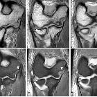
review of anatomy and common collateral ligament complex pathology using MRI. Posterolateral rotatory instability, stage 3B. Consecutive coronal T1-weighted MRI (a–c), and consecutive coronal FS PD-weighted MRI (d–f) showing an acute proximal full-thickness tear of the lateral ulnar collateral ligament and radial collateral ligament (white arrows) and anterior bundle of the medial collateral ligament complex sprain (white arrowheads)

The elbow:
review of anatomy and common collateral ligament complex pathology using MRI. Posterolateral rotatory instability, stage 3B. Consecutive coronal FS T1-weighted direct MR arthrographic images (a–f) showing a proximal partial-thickness tear of the anterior bundle of the medial collateral ligament complex (short arrows), proximal complete tear of the lateral ulnar collateral (long white arrows), and partial-thickness tear of the radial collateral ligament proper (white arrowhead)

The elbow:
review of anatomy and common collateral ligament complex pathology using MRI. Axial illustrations with superior view demonstrating the progression of the structures injured in the posterolateral rotatory instability

The elbow:
review of anatomy and common collateral ligament complex pathology using MRI. Anterior view of the elbow demonstrating the progression of the structures injured in a posterior dislocation, from lateral to medial. a Normal. b Proximal disruption of the lateral ulnar collateral ligament (white asterisk). c Proximal disruption of the lateral ulnar collateral ligament and the radial collateral ligament (black asterisk). d Anterior capsule tear (white arrow). Lateral ulnar collateral ligament (LUCL). Radial collateral ligament (RCL). Annular ligament (AL). Anterior bundle of the medial collateral ligament complex (A-MCL)

The elbow:
review of anatomy and common collateral ligament complex pathology using MRI. Posterolateral rotatory instability, stage 1. Coronal FS PD-weighted MRI (a, b) and coronal T1-weighted MRI (c, d) showing an avulsion of the proximal lateral ulnar collateral ligament and retraction of the osseous fragment (white arrows). Bone marrow edema is seen in the capitellum (white arrowheads) and radial head (white asterisks)

The elbow:
review of anatomy and common collateral ligament complex pathology using MRI. Posterolateral rotatory instability, stage 3B. Consecutive coronal FS T1-weighted direct MR arthrographic images (a, b), sagittal FS T1-weighted direct MR arthrographic image (c), and consecutive axial FS T1-weighted direct MR arthrographic images (d, e) showing a proximal complete tear of the lateral ulnar collateral ligament and radial collateral ligament proper (white short arrows), diffuse partial-thickness tear of the anterior bundle of the medial collateral ligament complex (white arrowheads), and a non-displaced fracture of the coronoid process (long white arrows)

The elbow:
review of anatomy and common collateral ligament complex pathology using MRI. Posterolateral rotatory instability, stage 3C. Consecutive coronal FS T1-weighted direct MR arthrographic images (a–c), and consecutive axial FS T1-weighted direct MR arthrographic images (d–f) demonstrating a full-thickness tear of the anterior bundle of the medial collateral ligament complex (yellow arrow), proximal full-thickness tear of the common flexor tendon (short white arrows), proximal full-thickness tear of the common extensor tendon (white arrowheads), proximal complete tear of the lateral ulnar collateral ligament and radial collateral ligament proper (long white arrows), and a complete tear of the posterior bundle of the medial collateral ligament complex (yellow arrowheads)

The elbow:
review of anatomy and common collateral ligament complex pathology using MRI. Posterolateral rotatory instability, stage 3B. Sagittal FS PD-weighted MRI (a) and consecutive coronal FS PD-weighted MRI (b, c) showing a posterolateral subluxation of the radial head (white arrowhead), proximal disruption of the lateral ulnar collateral ligament (white arrow), partial proximal disruption of the radial collateral ligament (yellow arrow), proximal disruption of the anterior bundle of the medial collateral ligament (blue arrow), radial head and posterior capitellar contusions (white asterisks), and joint effusion (black asterisks)

The elbow:
review of anatomy and common collateral ligament complex pathology using MRI. Posterolateral rotatory instability, stage 3B. Coronal T1-weighted MRI (a) and coronal FS PD-weighted MRI (b) showing an acute proximal common avulsion of the lateral ulnar collateral ligament and radial collateral ligament (white arrows), an acute avulsion of the anterior bundle of the medial collateral ligament complex (white arrowheads), a radial head fracture (white asterisks), a partial tear of the proximal common extensor tendon (yellow arrows), and joint effusion
Verletzungen des lateralen Kollateralbandkomplexes des Ellenbogens
Siehe auch:

 Assoziationen und Differentialdiagnosen zu Verletzungen des lateralen Kollateralbandkomplexes des Ellenbogens:
Verletzungen
des lateralen ulnaren Kollateralbandes am Ellenbogen
Verletzungen
des medialen Kollateralbandkomplexes des Ellenbogens


