benigne Tumoren des Magens

Großes
Leiomyom des Magens in der Doppelkontrastuntersuchung. Glatt begrenzter Tumor ohne Schleimhautdestruktion; häufigster submuköser Tumor im Magen; meist Zufallsbefund; kann jedoch auch ulzerieren und Ursache einer oberen GI-Blutung sein! Differentialdiagnostisch kommen in Frage: Leiomyoblastom, -sarkom, Lipom (negative Dichtewerte in der CT!), Hämangiom, Neurofibrom und Granular-Zell-Tumor, ektoper Pankreasrest, akutes Ulkus des Antrums, Kompression von außen durch vergrößerte Nachbarorgane oder Metastasen in diesen (Leber, Milz, Pankreas, Niere)

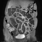
Invasive
inflammatory fibroid polyp of the stomach: a case report and literature review. Computed tomography (CT) findings. a Axial view. b Coronal view. c Sagittal view. CT image showing a hyper-enhancing tumor in the submucosa of the prepyloric antrum (arrows). CT showing a thickening of the muscularis propria layer and a hyper-enhancing lesion in the subserosa of the gastric antrum (arrow heads)

Gastric
lipoma presenting as a giant bulging mass in an oligosymptomatic patient: a case report. A computed tomography image before surgical exploration shows a large homogeneous mass with fat tissue density and central contrast captation with trabecular architecture (white arrow).


 Assoziationen und Differentialdiagnosen zu benigne Tumoren des Magens: