calcaneal fracture


Calcaneal fractures are the most common tarsal fracture and can occur in a variety of settings.
Epidemiology
The calcaneus is the most commonly fractured tarsal bone and accounts for about 2% of all fractures and ~60% of all tarsal fractures .
Pathology
Calcaneal fractures can be divided broadly into two types depending on whether there is articular involvement of the subtalar joint :
- anterior calcaneal process fracture
- calcaneal tuberosity avulsion fracture
- extra-articular body fracture
- medial sustentaculum
- intra-articular body fracture
The calcaneus is also a common site of stress fractures, occurring in the posterosuperior aspect.
Another method of classification is as
- type A fractures: the anterior process of the calcaneus is fractured
- type B: fracture of the mid calcaneus, trochlear process, and sustentaculum tali
- type C: fracture of the posterior tuberosity
Radiographic features
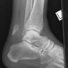
Plain radiograph
- Böhler's angle is the angle between two tangent lines drawn across the anterior and posterior borders of the calcaneus in the lateral view. When Böhler's angle becomes <20º it indicates a calcaneal fracture. On a lateral radiograph, an angle of Gissane of >130° suggests depression of the posterior facet of the subtalar joint.
- normal Böhler's angle does not exclude a fracture and is unreliable in children
- calcaneal stress fracture shows vertical linear sclerotic appearance
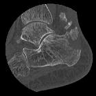
CT
CT is the modality of choice to evaluate calcaneal fracture. It can show the extent and extra- or intra-articular components of the fracture and hematoma along the sole of the foot (Mondor sign). Intra-articular fractures are often classified using the Sander classification system, which is one of the only systems that correlate well with patient outcome.
Practical points
If bilateral calcaneal fractures are seen, then the spine should also be evaluated for fracture as the mechanism of injury is often a large load to the axial skeleton, such as jumping from a second-story window.
If an intraarticular calcaneal fracture is seen, the images should be scrutinized for a lateral malleolar fleck sign (ankle), which raises the likelihood of peroneal tendon instability .
Siehe auch:
- Os calcaneus secundarius
- Böhlerwinkel
- calcaneal tuberosity avulsion fracture
- Fraktur Processus anterior calcanei
- Kalkaneusfraktur Klassifikation
- Kalkaneusfraktur Klassifikation Sanders
und weiter:


 Assoziationen und Differentialdiagnosen zu Kalkaneusfraktur: