Carpometacarpal bossing


Carpal boss
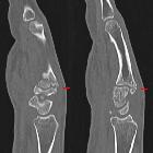
in der Computertomographie: 2 nebeneinander liegende Schichten einer sagittalen MPR.

Carpal boss
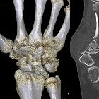
in der Computertomographie: VR-3D-Rekonstruktion. Pseudarthrotische Verbindung der Knochenvorsprünge am Metacarpale 3 und am Os trapezoideum.


Carpal boss
• Carpal boss - Ganzer Fall bei Radiopaedia

Carpal boss
• Carpal boss - Ganzer Fall bei Radiopaedia

Carpal boss
• CT carpal boss - Ganzer Fall bei Radiopaedia

Carpal boss
• Carpal boss - Ganzer Fall bei Radiopaedia

Carpal boss
• Carpal boss - Ganzer Fall bei Radiopaedia

Carpal boss
• Carpal boss - Ganzer Fall bei Radiopaedia

Carpal
bossing D3 mit Fraktur, möglicherweise nicht mehr ganz frisch, jedoch Klinik in den Bereich.

Pseudotumoural
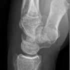
soft tissue lesions of the hand and wrist: a pictorial review. Carpal boss. a Plain radiograph (lateral view) showing a bony prominence at the dorsal aspect of the carpometacarpal joint (arrow). b Sagittal fat-suppressed (FS) TSE T2-weighted image (WI). Note bone marrow oedema and subchondral cyst formation at the carpo-metacarpal joint (arrows)
The carpal boss is a hypertrophied bony protuberance on the dorsal surfaces of the base of the second or third metacarpals, near the capitate and trapezium. It may be bilateral.
Pathology
The condition may represent one or more of:
- degenerative osteophyte formation
- os styloideum (an accessory ossicle of the wrist)
- bony prominence at the base of the second or third metacarpals (which is often called "styloid process")
Pain may be the result of a ganglion, inflamed bursa or extensor tendon slipping over the bony prominence.
Radiographic features
The radiographic appearance of a carpal boss is characteristic, however optimal visualization of the boss may be difficult to obtain due to superimposed bony structures.
CT may show degenerative disease at a pseudoarthrosis, and MRI may show edema related to abnormal motion.
Siehe auch:

 Assoziationen und Differentialdiagnosen zu Carpal boss:
pseudotumoröse
Weichteilläsionen von Hand und Handgelenk


