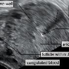
cervical ectopic pregnancy


Cervical ectopic pregnancy is a rare subtype of ectopic pregnancy, in which a gestational sac is seen at the endocervical canal below the closed internal os.
Epidemiology
It accounts for ~0.15-1% of all ectopic pregnancies.
Pathology
Implantation of the fertilized ovum occurs within the cervix rather than the uterine cavity. Unless the fetal heart rate can be identified, it is difficult to distinguish from a miscarriage with fetal parts in the cervical os.
Risk factors
- variant anatomy
- fibroids
- IUCD
- Asherman syndrome
- IVF
- repeated dilatation and curettage
Radiographic assessment
Ultrasound
Can be seen as a gestational sac within the distended cervix which gives an hour-glass appearance to the uterus. Usually, internal os is closed. At times the gestational sac extends into the lower uterine segment (abnormally low sac position). There is hyperechoic decidual reaction around the gestational sac.
Color Doppler imaging can be helpful in showing a hypervascular trophoblastic ring in the cervical region in cases of live cervical ectopic pregnancies.
Treatment and prognosis
The goal is to treat the condition whilst minimizing the risk of severe hemorrhage, and to preserve the patient's future reproductive potential.
Medical management options include methotrexate (a folate antagonist) either administered systemically or by direct injection, or potassium chloride (direct injection).
Surgical curettage runs the risk of life-threatening hemorrhage. Preoperative uterine embolization is an option for reduction of hemorrhage risk.
Complications
Severe hemorrhage is the main complication of surgical management. It may result in the need for hysterectomy, and is potentially life-threatening.
Differential diagnosis
- it is important to consider miscarriage in progress as a differential when the gestational sac is located within the endocervical canal; features suggestive of a miscarriage include:
- absent embryonic cardiac activity
- open internal os: lack of typical hourglass appearance
- sac shape and location often changes on serial scans and it may be possible to alter the location of the sac with gentle probe maneuvering ("sliding sac sign")
- subsequent loss of the fetal sac on a repeat ultrasound confirms miscarriage
Siehe auch:
und weiter:


 Assoziationen und Differentialdiagnosen zu