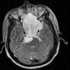
Chordom am Clivus

Combined use
of maxillomandibular swing approach and neurosurgical ultrasonic aspirator in the management of extensive clival chordoma: A case report. Sagittal and axial views of brain MRI scan. Image shows tumour in the nasopharynx extending from nasal cavity to brainstem posteriorly.

Radiological
review of skull lesions. Chordoma. Axial head CT (a) shows an osteolytic destructive lesion involving the clivus (thick arrows) with extension into the posterior sphenoid sinus (arrowhead) and impression on the pons (curved arrow). Axial T1-weighted (b), axial T2-weighted (c) and sagittal post-contrast T1-weighted (d) images demonstrate an expansile mass centred at the clivus extending into the sphenoid sinus (arrowhead), left cavernous region (short, thick arrows) and partially encasing the left internal carotid artery. This mass enhances and displaces the pituitary gland (thin, long arrow) and has mass effect on the left aspect of the pons (curved arrow)

Clival
chordoma - CT and MRI. Infiltrative clival lesion with intralesional calcifications, some chondroid-like (dots-&-commas). Invades posterior nasopharynx, carotid canals (a), and endocranium, bulging and "thumbprinting" the anterior aspect of the pons (b). Also invades both cavernous venous sinuses (b).

Clival
chordoma - CT and MRI. Fig 1c. CECT shows heterogeneous, honeycomb-like enhancement of the clival lesion (c).

Clival
chordoma - CT and MRI. Axial T1SE. Homogeneously intermediate-hypointensity. Right carotid artery encasement.

Clival
chordoma - CT and MRI. Coronal T2TSE. Heterogeneously hyperintense with cystic foci.

Clival
chordoma - CT and MRI. Sagittal T1+contrast. Heterogeneous “honeycombing” enhancement, with a predominantly peripheral distribution. Endocraneal invasion is evident, with pontine "thumbprinting".

Clival
chordoma - CT and MRI. DWI. Discrete hyperintensity of the lesion.

Clival
chordoma - CT and MRI. ADC map. Region of interest (red circle) relatively low ADC values (615E-6 +/- 65E-6 mm^2/s).
 Assoziationen und Differentialdiagnosen zu Chordom am Clivus:


