Ecchordosis physaliphora


Ecchordosis physaliphora is a congenital benign hamartomatous lesion derived from notochord remnants, usually located in the retroclival prepontine region, but can be found anywhere from the skull base to the sacrum.
Terminology
There has been some controversy as to whether intradural chordoma and large ecchordosis physaliphora are different entities. Some authors (such as Wolfe et al.) proposed the name 'intradural chordoma' for all intradural notochordal remnant lesions . Others (such as Rodriguez et al.) proposed that all intradural notochordal remnant lesions should be called ecchordosis physaliphora, until chordoma are pathologically proven to arise from the intradural compartment . However, they are currently considered distinct pathologies with a common origin.
Clinical presentation
Unlike chordomas which are often symptomatic due to brainstem or cranial nerve compression, patients with ecchordosis physaliphora are usually asymptomatic. They are found in ~2% of autopsies .
Pathology
Ecchordosis physaliphora arise from remaining notochord cells along the axis of the spine after embryogenesis. Unfortunately, ecchordosis physaliphora and chordoma are histologically indistinguishable, other than by examining the margins, the latter demonstrating infiltrative growth.
Radiographic features
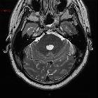
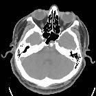
CT
CT is generally not sensitive for such lesions, mainly because of posterior fossa artifacts and the near CSF density of the mass. The bony clival defect is, however, visible as a well-demarcated smoothly corticated region, without aggressive features.
Occasionally an osseous stalk is seen at the base of the lesion which is said to be pathognomonic in this context .
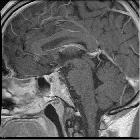
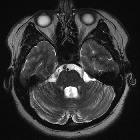
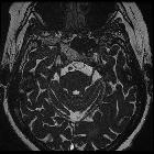
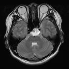
MRI
A stalk-like connection to the clivus is usually seen if high-resolution images are obtained.
Apart from the characteristic location (retroclival, prepontine, and intradural), MRI findings are non-specific, with signal similar to CSF:
- T1: hypointense
- T2: hyperintense
- T1 C+ (Gd): variable, however, most cases have not shown substantial enhancement
History and etymology
Hubert von Luschka (1820-1875), a German pathologist, first described the finding of pathologic ectopic notochordal tissue at the posterior clivus in 1856.
Differential diagnosis
The differential diagnosis of retroclival intradural lesions consists mainly of :
- chordoma: generally symptomatic and enhances after contrast administration
- benign notochordal cell tumor (BNCT)
- skull base metastasis
- dermoid cyst
- epidermoid cyst
- arachnoid cyst: communicates with subarachnoid space, follows CSF intensity pattern and no enhancement
Siehe auch:
- Raumforderungen des Clivus
- Dermoidzyste
- epidermale Inklusionszyste
- Metastase
- Canalis basilaris medianus
- Chordom am Clivus
- zystische Läsion im Klivus
- intrakranielle neuroenterische Zyste
- Foveola pharyngica
- Venae diploicae im Clivus
und weiter:


 Assoziationen und Differentialdiagnosen zu Ecchordosis physaliphora:


