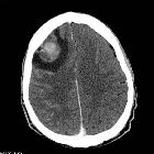
choriocarcinoma of the uterus


Uterine choriocarcinomas are one of the commonest choriocarcinomas and are often associated with gestational trophoblastic disease (GTD).
Epidemiology
These tumors typically occur in women of childbearing age as a gestational choriocarcinoma. Most such cases present within one year of an antecedent pregnancy . Presentation beyond reproductive age as a non-gestational choriocarcinoma is a rare occurrence .
Pathology
As with choriocarcinomas in general, it is a highly vascular neoplasm. The tumor comprises of trophoblastic cells. On histology, there is an absence of chorionic villi (c.f. other forms of GTD).
Etiology
In the case of gestational choriocarcinoma, approximately 5% of cases of complete hydatidiform mole are followed by choriocarcinoma.
Only about half the cases of choriocarcinoma arise from a complete hydatidiform mole. An additional 25% of cases arise after normal pregnancies, and 25% follow spontaneous abortion or ectopic pregnancy.
Staging
Radiographic features
Regardless of the imaging modality used, choriocarcinoma often appears as a mass enlarging the uterus. Sometimes it manifests as a discrete, central, infiltrative mass. Its heterogeneous appearance correlates with necrosis and hemorrhage that characterize these lesions.
Ultrasound
They can have variable sonographic appearances, but customarily present as an infiltrative heterogeneous mass invading myometrium and beyond. The uterus may be enlarged. Cystic areas from necrosis and hemorrhage may be present.
The ovaries can be shown enlarged due to cysts secondary to increased levels of β-hCG (see case 3).
Treatment and prognosis
Those cases that arise after complete hydatidiform mole are nearly all cured by chemotherapy (in contrast to the others, which have a less favorable prognosis).
Siehe auch:
- gestational trophoblastic disease
- Chorionkarzinom
- complete hydatidiform mole
- gestational choriocarcinoma
- non-gestational choriocarcinoma
- Chorionkarzinom Staging
und weiter:

 Assoziationen und Differentialdiagnosen zu Chorionkarzinom des Uterus:


