chronic recurrent multifocal osteomyelitis


Chronic recurrent multifocal osteomyelitis (CRMO) is an idiopathic inflammatory bone disorder seen primarily in children and adolescents. It is often a diagnosis of exclusion once underlying infection and neoplasia have been ruled out. However, there are some cases in which lesion location and morphology combined with clinical assessment can yield a diagnosis without biopsy.
Epidemiology
There is a female predominance, with 85% of cases reported in women.
Clinical presentation
The typical clinical picture of CRMO is a child or young adult with a history of chronic multifocal bone pain. Systemic symptoms of fever, weight loss or lethargy are uncommon but can occur. Biopsy of osseous lesions shows an inflammatory reaction with failure to culture any organisms.
Pathology
The etiology and pathogenesis of CRMO remains unclear. While early reports suggested an infective etiology, no consistent pathogen has been demonstrated in subsequent studies and biopsy cultures are usually negative.
As the condition can be associated with skin conditions, like psoriasis, or inflammatory bowel disease an autoimmune cause is thought likely.
Radiographic features
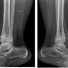
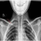
The most common sites of disease are the metaphyses or metaphyseal equivalents of the long bones, most frequently in the tibia. However, a wide range of skeletal site involvement has been reported.
Clavicle involvement is a characteristic finding since this is an uncommon location for haematogenous osteomyelitis. However, imaging findings overall remain suggestive rather than pathognomonic for CRMO.
Plain radiograph
Initially to examine the symptomatic site:
- early stages: osteolytic lesion
- later stages: progressive sclerosis
Lesions can range between purely osteolytic, osteolytic with a sclerotic rim, mixed lytic and sclerotic, and purely sclerotic.
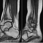
MRI
Useful for determining the extent of disease with whole-body STIR, including disease sites that may be clinically occult and can be used for subsequent follow-up. Findings can include:
- bone marrow edema
- periostitis
- soft tissue edema
- extension across the physis
- areas of post-gadolinium enhancement
Findings that should raise the possibility of haematogenous osteomyelitis over CRMO include:
- large fluid collection
- abscess
- fistulous tract
- bony sequestrum
Nuclear medicine
Similar to whole-body MRI, bone scintigraphy demonstrates multifocal uptake in keeping with the multifocal nature of the disease. In children, MRI is preferred to avoid radiation dose.
Treatment and prognosis
CRMO should be treated by a rheumatologist. While non-steroidal anti-inflammatory drugs may cause some symptomatic relief, there is often the need for a greater degree of immunosuppression and possibly biological agents. These are often similar drugs that are used in the treatment of juvenile arthritis and the associated multi-disciplinary rheumatology team is vital to ensure adequate physiotherapy input.
Long-term clinical follow up with whole-body MRI may be needed until the patient is in radiological remission or a steady-state is achieved.
History and etymology
The syndrome of CRMO was originally described in 1972 in four children under the name “subacute and chronic recurrent osteomyelitis'' . The term chronic recurrent multifocal osteomyelitis was first used by Probst et al.
Differential diagnosis
- infectious osteomyelitis (subacute and chronic)
- Langerhans cell histiocytosis
- Ewing sarcoma
Siehe auch:
und weiter:


 Assoziationen und Differentialdiagnosen zu Chronisch rekurrierende multifokale Osteomyelitis: