costal cartilage fracture


Costal cartilage fractures are fractures of the cartilage connecting the ribs anteriorly to the sternum.
Epidemiology
There is little published data on costal cartilage fractures. Most reported cases are in males and resulted from blunt trauma or a fall .
Clinical presentation
In young children, a costal cartilage fracture can present as a chest wall mass associated with pain (differentials include neoplasm and post-traumatic hematoma). In older patients, pain is the primary complaint .
Pathology
Costal cartilages can fracture due to blunt trauma sustained in high-energy trauma or a fall. While the more immobile first and second ribs seem to be more prone to costochondral separation, the lower ribs can suffer costal cartilage fracturing more easily .
Disruptions or fractures of costal cartilage might result in an unstable rib cage and may expose thoracic contents, such as the heart, to injury .
Radiographic features
Plain radiography
Fractures of the costal cartilage are challenging to establish on physical examination and not visible on plain radiographs unless there is severe calcification of the cartilage . Due to their low incidence, they may also be easily overlooked on additional imaging.
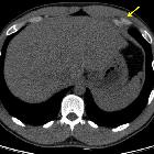
CT
CT is a reliable method of identifying costal cartilage fractures . They tend to be mid-substance (i.e. in the center of the costal cartilage), and CT offers the advantage of demonstrating other injuries, such as rib fractures and involvement of lung parenchyma.
Ultrasound
Ultrasound can be used reliably to diagnose costal cartilage fractures and can increase the sensitivity of their detection when used together with CT scanning . Unlike CT it does not involve ionizing radiation and is, therefore, preferable in a pediatric population and for follow-up examinations . Furthermore, it may be helpful to perform an ultrasound examination when there is a high clinical suspicion, with or without a mass, and other modalities have not demonstrated an injury .
Signs of a cartilage fracture include a fracture line, disruption of the anterior echogenic margin, a step-off deformity or gas located at the costochondral junction .
Pitfalls include :
- confusion of the pleural surface with rib cortex
- misinterpretation as a fracture due to:
- transducer overlying a rib and either an intercostal space or scapula
- costal cartilage calcifications parallel to the rib surface (less likely in pediatric population)
MRI
MRI can demonstrate costal cartilage fractures as well and, like ultrasound, lack exposure to ionizing radiation.
Treatment and prognosis
It is unclear if costal cartilage fractures completely heal , but reported cases state return to normal and sports activities when the pain goes away . It is not known if the supposedly healed cartilage can sustain the same loads as the original cartilage. When returning to sports, especially contact sports at risk for direct trauma (e.g. hockey, rugby), protective padding may be used for an additional period as a precaution to allow for further healing while reducing the risk of any repeated injury.
Siehe auch:
- Rippenfraktur
- sternocostale Luxation
- Fraktur des Rippenknorpels der ersten Rippe
- Rippenknorpel Anomalie
- Verletzungem am sternokostalen Übergang
und weiter:


 Assoziationen und Differentialdiagnosen zu Fraktur Rippenknorpel:
