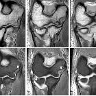
CT Arthrographie Ellenbogen

Arthrografie
und CT-Arthrografie Ellenbogen mit Radiusköpfchenfraktur und Abscherung eines chondralen Flakes am Capitulum humeri: Die Injektion erfolgte von dorsal (ca. 4 ml). Der Knorpelüberzug ist gegen das röntgendichte Kontrastmittel gut abgrenzbar.

The elbow:
review of anatomy and common collateral ligament complex pathology using MRI. Sagittal FS T1-weighted direct MR arthrographic images (a, b), coronal FS T1-weighted direct MR arthrographic image (c), and axial FS T1-weighted direct MR arthrographic images (d–f) showing a proximal partial-thickness proximal tear of the anterior bundle of the medial collateral ligament complex (short arrow), disruption and stripping of the posterior and lateral capsular structures (white asterisks), intra-articular bodies (long arrows), and full-thickness defect at the posterior joint line (arrowhead). Note ulnohumeral incongruity (a) and radiocapitellar incongruity (b)

Overuse-related
instability of the elbow: the role of CT-arthrography. In-plane US-guided contrast agent injection using a posterior access. The spinal needle is guided through the triceps tendon, with an approximate 30° angle between the needle and the probe in order to obtain an optimal visualization of the needle tip advancing to the posterior articular recess

Overuse-related
instability of the elbow: the role of CT-arthrography. In-plane US-guided intra-articular injection (a). The spinal needle is clearly visible as a hyperechoic linear image, as the tip (white arrow) approaches the designed target. The posterior articular recess (asterisk) expands as contrast agent is injected, becoming visible as a hypoechoic sac, confirming the appropriateness of the procedure (b)

Overuse-related
instability of the elbow: the role of CT-arthrography. The patient is positioned prone on the CT scanning table, with the affected upper limb elevated over the head and with a 45° flexion of the elbow

Overuse-related
instability of the elbow: the role of CT-arthrography. CT-arthrography coronal images (a–d) and axial images (e–h) belonging to four different patients, three of which display different aspects of ligament derangement involving the A-MCL (white arrows). Intact anterior band (A-MCL) of the medial collateral ligament (MCL) complex (a, e and b, f). Widening of the medial aspect of the articular recess, associated to a “bow-shaped” loose A-MCL, with no evidence of fiber tearing, suggestive of ligament laxity (b, f). Thickened and irregular A-MCL, showing a partial-thickness tear (c, g) and a full-thickness tear (d, h); contrast agent progressively spills through the ligament, as fibers are gradually disrupted as a consequence of micro-tearing and damage progresses from a partial to a complete tear

Overuse-related
instability of the elbow: the role of CT-arthrography. Professional tennis player with micro-tearing of the anterior band (A-MCL). Oblique coronal reformatted images depicting the A-MCL along its longitudinal axis, up to its insertion at the sublime tubercle (ST), highlight a thickened ligament (asterisk) with multiple strands of contrast enhancement within its fibers, due to micro-tearing (white arrows)

Overuse-related
instability of the elbow: the role of CT-arthrography. Professional tennis player with chronic medial elbow pain, exacerbated during serve, not responsive to medical and physical treatments. MRI coronal fat-suppressed T2-weighted images (a) show a high-intensity signal at the anterior bundle (A-MCL), suggestive of partial tear (black arrows). CT-arthrography reformatted images along the major axis of the anterior bundle (b) demonstrate a thickened but continuous ligament (white arrows). US scan prior to contrast agent injection (c) confirms diagnosis by showing a thickened, hypoechoic ligament with irregular margins, although regularly inserted (yellow arrows)

Overuse-related
instability of the elbow: the role of CT-arthrography. CT-arthrography sagittal (a) and axial (b–d) images of two different patients with valgus extension overload (VEO) syndrome, showing osteophyte formation (b, c) over the medial aspect of the olecranon (white arrows) and an intra-articular loose body (d) located medially at the posteromedial recess (green arrow). Synovial thickening associated to synovitis (a, b) is also noticeable (yellow arrows)

Overuse-related
instability of the elbow: the role of CT-arthrography. Posterolateral rotatory instability (PLRI) sagittal, coronal and axial images (a–c). Full-thickness tear of the lateral collateral ligament (LCL) complex (white arrows) with extra-articular leakage of iodinated contrast medium within fibers of the common extensor tendon (asterisks)

Overuse-related
instability of the elbow: the role of CT-arthrography. CT-arthrography coronal (a, c, e, g) and axial (b, d, f, h) images belonging to four patients, three of which display different aspects of symptomatic minor instability of the lateral elbow (SMILE), involving the RCL (white arrows). Intact radial band (RCL) of the lateral collateral ligament (LCL) complex (a, b). Widening of the lateral aspect of the articular recess associated to a curve-shaped, loose RCL with no evidence of fiber tearing, suggestive of patholaxity (c, d). Thickened and irregular RCL showing partial-thickness tear (e, f) and full-thickness tear (g, h); iodinated contrast progressively leaks through the ligament, as fibers are gradually disrupted and damage progresses from a partial to a full-thickness tear

Overuse-related
instability of the elbow: the role of CT-arthrography. CT-arthrography sagittal (a, d, g), axial (b, e, h) and coronal (c, f, i) images belonging to three patients, two of which display different SMILE stages involving the AL (white arrows). Intact annular ligament (AL) of the lateral collateral ligament (LCL) complex (a–c). Loose AL with no evidence of fiber tearing, displaying the “loose collar sign”, suggestive of patholaxity (d–f). Thickened and irregular AL showing a partial-thickness tear (g–i); ligament fibers appear delaminated as iodinated contrast permeates through their layers

Overuse-related
instability of the elbow: the role of CT-arthrography. Symptomatic minor instability of the lateral elbow (SMILE) with a high-degree lesion of the annular ligament (AL) of the LCL complex. Consecutive sagittal (a, b), coronal (c, d) and axial (e, f) CT-arthrography scans highlight the ruptured AL (white arrows) and display extra-articular leakage (asterisks) through the damaged articular capsule
CT Arthrographie Ellenbogen
Siehe auch:

 Assoziationen und Differentialdiagnosen zu CT Arthrographie Ellenbogen:


