diskoligamentäre Verletzungen der Wirbelsäule

Minimally
displaced unilateral facet fracture of cervical spine can lead to spinal cord injury: a report of two cases. Magnetic resonance imaging using a sagittal short T1 inversion recovery (STIR) sequence and b axial T2 weighted image at C5/6 at re-admission shows disc injury and spinal cord compression by a posterior epidural mass accompanied by an intramedullary signal intensity change at this level. Prevertebral soft-tissue edema, injury of the interspinous ligament, and a narrowed canal also are evident. There is no flow void in right vertebral artery on axial T2 weighted image, suggesting vertebral artery injury
Minimally
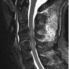
displaced unilateral facet fracture of cervical spine can lead to spinal cord injury: a report of two cases. Magnetic resonance imaging using sagittal short T1 inversion recovery (STIR) sequence showed an intramedullary signal change at C4/5 with disc injury. Prevertebral soft-tissue edema and injury of the interspinous ligament, and narrowed canal also are evident

Cervical
Spine MRI of patient with SCI: C4 fracture and dislocation, spinal cord compression
diskoligamentäre Verletzungen der Wirbelsäule
Siehe auch:


 Assoziationen und Differentialdiagnosen zu diskoligamentäre Verletzungen der Wirbelsäule:

