duplex collecting system


A duplex collecting system, or duplicated collecting system, is one of the most common congenital renal tract abnormalities. It is characterized by an incomplete fusion of upper and lower pole moieties resulting in a variety of complete or incomplete duplications of the collecting system. While considered an anatomical variant, duplex collecting systems may be complicated by vesicoureteric reflux, obstruction or ureterocele.
Epidemiology
Duplex collecting systems are seen in 0.7% of the healthy adult population and 2-4% of patients investigated for urinary tract symptoms .
Clinical presentation
Most duplicated systems are asymptomatic and diagnosed incidentally. However, where symptoms do occur (infection, reflux or obstruction), the patient is likely to have completely duplicated ureters. Occasionally, hydronephrosis can be severe enough to result in flank discomfort or even a palpable mass.
Pathology
Embryologically, duplication occurs when two separate ureteric buds arise from a single Wolffian duct. Interestingly, and explaining the Weigert-Meyer rule, the future lower pole ureter separates from Wolffian duct earlier and thus migrates superiorly and laterally as the urogenital sinus grows.
Duplication can be variable. At one end of the spectrum, there is merely a duplication of the renal pelvis, draining via a single ureter. At the other extreme, two separate collecting systems drain independently into the bladder or ectopically (see below).
Duplex systems may be unilateral or bilateral and can be associated with a variety of other congenital abnormalities of the urinary tract, e.g. ureterocele.
Associations
Classification
Duplex collecting system or duplex kidney anomalies can be classified into the following categories depending on the level or lack of fusion :
- duplex kidney: two separate pelvicalyceal systems draining a single renal parenchyma
- duplex collecting system: a duplex kidney draining into:
- single ureter: i.e. duplex kidney's duplication pelvicalyceal systems uniting at the pelviureteric junction (PUJ)
- bifid ureter (ureter fissus): two ureters that unite before emptying into the bladder
- double ureter (complete duplication)
- bifid collecting system: refers to a duplex kidney with the two separate pelvicalyceal collecting systems uniting at the PUJ or as bifid ureters
- double/duplicated ureters (or collecting system): two ureters that drain separately into the bladder or genital tract
Radiographic features
As the abnormality is an anatomic alteration, all modalities able to image the renal tract may be able to visualize the typical features.
General features include:
- duplicated ureters extending a variable distance down to the bladder
- obstruction of the upper pole moiety down to the bladder, often with a ureterocele
- vesicoureteric reflux into the lower pole moiety, often due to distortion in its insertion by the aforementioned ureterocele
- ectopic insertion of the upper pole moiety e.g. into the prostatic urethra in males or vaginal vault in females
Additionally, if reflux is significant, evidence of reflux nephropathy may be evident.
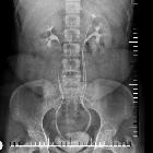
Fluoroscopy
Excretory urography (IVP)
Although able to elegantly image both collecting systems, one should be aware that a poorly functioning system may not excrete contrast. In such a situation, the functioning lower pole moiety will be inferiorly displaced, taking on the so-called "drooping lily appearance" . The differential for such an appearance is that of an upper pole mass or cyst.
Ultrasound
Ultrasound, when no obstruction/hydronephrosis is present can be suboptimal in the detection of a duplicated system and will especially struggle to distinguish between partial and complete duplication. It is certainly able to detect ureteroceles, if present.
It provides excellent anatomic information but does not necessarily differentiate a bifid renal pelvis from a bifid ureter or two complete ureters.
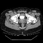
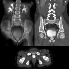
CT
CT can delineate all abnormalities essentially, especially when performed during the excretory (IVP) phase (contrast outlining the collecting systems). CT reconstruction software can produce striking single images of the collecting systems. In an unobstructed system, the diagnosis can be difficult. A duplicated renal collecting system can be suspected by identifying the so-called faceless kidney.
Nuclear medicine
Renal scintigraphy is significantly impaired in its ability to identify non-obstructed systems, as the spatial resolution is poor. However, it is able to evaluate renal function and is particularly useful in planning corrective surgery.
Treatment and prognosis
A duplex kidney usually does not require any treatment per se, however, complications may necessitate intervention:
- vesicoureteric reflux into lower pole moiety
- marked hydronephrosis of the upper pole moiety may have a mass effect or become infected
Siehe auch:
- Ureterozele
- wolffian duct
- Vesikoureteraler Reflux
- Hydronephrose
- Fanconi-Anämie
- faceless kidney
- developmental anomalies of the kidney and ureter
- Weigert-Meyer Regel
- drooping lily appearance
und weiter:
- genitourinary curriculum
- Bertini-Säulen
- junktionaler Parenchymdefekt der Niere
- fetal pyelectasis
- Malrotation der Niere
- Akrorenales Syndrom
- Anomalien des urogenitalen Systems
- Doppelniere mit Hydronephrose der oberen Anlage durch Ureterektopie
- developmental renal anomalies
- Doppelniere Malrotation
- ektope Uretermündung
- kongenitaler Megaureter
- drooping lily sign
- Entwicklungsanomalien der Niere


 Assoziationen und Differentialdiagnosen zu Doppelniere:


