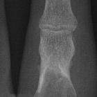
enchondroma vs low grade chondrosarcoma

Is PET–CT
an accurate method for the differential diagnosis between chondroma and chondrosarcoma?. Example of an MRI image of the shoulder, where it is not possible to confirm whether the lesion is a chondroma or a chondrosarcoma. In a axial T2 MRI image of the proximal humerus, and b T1 image. In c and d, the bone lesion can be visualized in T2 and T1 images. In e, axial PET and in f coronal images show the proximal region of the left humerus. Note the PET–CT presenting the volumetric region of interest (VOI) with an SUVmax = 2.0
Distinguishing between enchondromas and low-grade conventional chondrosarcomas is a frequent difficulty as the lesions are both histologically and radiographically very similar.
It is important to remember, though, that differentiating between them may be a moot point since both can either be closely followed up clinically and radiologically or treated if symptomatic.
Radiographic features
Useful features include:
- size
- lesion size over 5-6 cm favors chondrosarcomas
- cortical breach
- seen in 88% of long bone chondrosarcomas
- seen in only 8% of enchondromas
- deep endosteal scalloping involving > 2/3 of cortical thickness
- seen in 90% of chondrosarcomas
- seen in only 10% of enchondromas
- permeative or moth-eaten bone appearance
- seen in high-grade chondrosarcomas, not in low-grade tumors
- soft tissue mass beyond bone
- not seen in enchondroma
- increased uptake on bone scan
- seen in 82% of chondrosarcomas
- seen in only 21% of enchondromas
- location
- hands and feet are uncommon locations for chondrosarcoma
- outside hand and feet, chondrosarcomas outnumber enchondromas 5:1
- spine, pelvis, sacrum, and ribs are rare locations for enchondromas
- patient age
- enchondromas commonly appear in young adults
- chondrosarcomas tend to appear in middle-aged patients
- pain
- chondrosarcomas almost always present with pain
- enchondromas are painless unless they cause a pathological fracture
Siehe auch:
und weiter:

 Assoziationen und Differentialdiagnosen zu Unterscheidung Enchondrom Chondrosarkom:

