enchondroma


Enchondromas, also known as chondromas, are relatively common intramedullary cartilage neoplasms with benign imaging features. They share histologic features with low-grade chondrosarcoma, and are sometimes classified under the umbrella term low-grade chondral series tumors.
Enchondromas account for the 'E' in the popular mnemonic for lytic bone lesions FEGNOMASHIC.
Epidemiology
- most frequently diagnosed in childhood to early adulthood with a peak incidence of 10-30 years
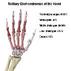
- most common primary benign bone tumor of hand/wrist
- account for ~5% (range 3-10%) of all bone tumors, and ~17.5% (range 12-24%) of benign bone tumors
Associations
Two syndromes are associated with multiple enchondromas:
Clinical presentation
Enchondromas are most commonly an incidental finding, most significant in that they shouldn't be confused with more aggressive lesions.
As a rule, enchondromas should be asymptomatic; however, lesions of the hands/feet may present with pain from pathological fracture or impending fracture.
Malignant transformation into low-grade chondrosarcoma is rare and may present with pain.
Pathology
Enchondromas comprise lobules of mature hyaline cartilage which are partially or completely encased by surrounding normal bone . The cartilaginous lobules may undergo endochondral ossification, often resulting in the characteristic 'rings and arcs' pattern of mineralization.
They arise from rests of growth plate cartilage/chondrocytes which become isolated within mature bone. Hence, they may be seen in any bone formed from cartilage.
By definition, they show no histologic evidence of local invasion (which would suggest low-grade chondrosarcoma). However, it is important to be aware that enchondroma cannot be reliably distinguished from chondrosarcoma by histology, and diagnosis depends on correlation of clinical, imaging, and pathology findings .
Grossly, lesions are usually <3 cm, translucent, nodular, and are grossly grayish-blue.
Location
Enchondromas are typically located in a central or eccentric position within the medullary cavity of tubular bones:
- small tubular bones of the hands and feet (~50%)
- proximal phalanx most common
- large tubular bones
- e.g. femur, tibia, humerus
- rare: (consider chondrosarcoma)
- pelvis
- ribs
- scapula
Rarely an enchondroma may extend through the cortex and demonstrate an exophytic growth pattern. This is known as an enchondroma protuberans, and may either be seen sporadically or as part of Ollier disease .
Radiographic features
Enchondromas have a somewhat variable appearance by imaging, although characterization by excluding suspicious features is key. Since most are asymptomatic incidental findings, lesions in a characteristic location and appearance are not usually further investigated.
Imaging is generally less helpful in corroborating benignity of lesions in the hands/feet, as well as in enchondromatosis or skeletally immature patients .
Radiograph and CT
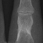
Enchondromas have a variable appearance, although typically they are small <5 cm lytic lesions with non-aggressive features:
- narrow zone of transition
- sharply defined margins
- +/- chondroid calcification (rings and arcs calcification)
- often no matrix mineralization (purely lytic) in the hands/feet
- +/- expansile
- more commonly in hands/feet
- may have mild endosteal scalloping
- should not "grow" through cortex (unless pathologic fracture)
- pertinent negatives :
- no gross bone destruction
- no periosteal reaction
- no soft tissue mass
The majority of enchondromas more frequently arise in the metaphyseal region, owing presumably to their origin from the growth plate , although they are frequently seen in the diaphysis. They only rarely are seen in the epiphysis, and a cartilaginous lesion in an epiphysis is more likely to be a chondrosarcoma .
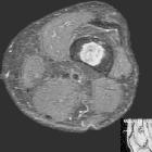
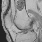
MRI
MRI is useful in evaluating soft tissue extension and for confirming the diagnosis. Enchondromas appear as well-circumscribed somewhat lobulated masses replacing marrow .
- T1
- intermediate to low-signal
- internal foci of low signal of “rings and arcs” characteristic of a chondroid matrix
- T1 C+ (Gd)
- enhancement is variable and may be seen both peripherally or of translesional septae
- similar pattern of enhancement may be seen in chondrosarcomas
- T2
- sharply defined
- predominantly high signal
- internal foci of low signal of “rings and arcs” characteristic of a chondroid matrix
- no bone marrow or soft tissue edema
Descriptions should include
- location i.e. central or peripheral
- presence and amount of endosteal scalloping
Differentiation of an enchondroma from low-grade chondrosarcoma is problematic, as they can have similar appearances. See enchondroma vs low-grade chondrosarcoma.
Nuclear medicine
Increased uptake on the bone scan can be seen with enchondromas. Intense uptake occurs with an underlying pathological fracture or cortical expansion in small bones .
Treatment and prognosis
The majority of enchondromas remain asymptomatic and require no treatment.
Pathologic fractures are commonly treated by curettage and bone grafting, with follow-up x-rays to monitor for healing and recurrence. An incisional biopsy is obtained intraoperatively. Recurrence is reported in 2-15% and suggests malignancy .
If malignant transformation is suspected, which occurs in less than 5% of cases, then treatment is more aggressive .
Complications
- pathological fracture
- malignant transformation into chondrosarcoma
Differential diagnosis
The differential is significantly affected by the modality in question, and most entities below can be excluded with MRI. The exception is chondrosarcoma.
- bone infarct
- chondrosarcoma
- difficult to distinguish
- see: enchondroma vs low grade chondrosarcoma
- intraosseous ganglion
- other benign lytic bone lesions
- lytic metastasis to bone
- granulomatous disease
Siehe auch:
- Tuberkulose
- Sarkoidose
- Chondrosarkom
- kartilaginäre Exostose
- Osteolyse Fingerknochen
- intraossäres Ganglion
- MRT Enchondrom
- Ollier-Syndrom
- Knocheninfarkt
- Chondroblastom
- benigne Osteolysen
- Enchondrom proximaler Humerus
- Chondrom
- pathologische Fraktur bei Enchondrom
- bizarre parosteale osteochondromatöse Proliferation
- Unterscheidung Enchondrom Chondrosarkom
- Maffucci-Syndrom
- Enchondrom in der Skelettszintigraphie
- Chondromatose
- Enchondrom des Mittelfußknochens
- Enchondrom Verteilung der Lokalisation
- Verteilung von Enchondromen an der Hand
- Enchondrom Phalangen
und weiter:
- endosteal scalloping
- Enchondrom Femur
- Enchondrom Humerus
- Riesenzelltumor
- popkornartige Verkalkungen
- Enchondrom Mittelhandknochen
- Türflügelzeichen
- radiologisches muskuloskelettales Curriculum
- Riesenzelltumor des Knochens
- Knochentumoren
- Enchondroma protuberans
- epiphysäre Knochentumoren
- osseous lesions preferentially involving the epiphysis
- Enchondrom distales Femur
- diaphyseal lesions (mnemonic)
- solitary sclerotic bone lesion
- Ostitis fibrosa cystica
- ring- und bogenförmige Verkalkungen
- lytic bone lesion (mnemonic)
- Osteoklastom
- Knochenläsionen der Epiphyse
- Metachondromatose
- Enchondrom der Rippen
- focal sclerotic bony lesions (mnemonic)
- multiple lytic bone lesions (mnemonic)
- lytic rib lesion (mnemonic)
- verkalktes Enchondrom
- Läsionen der Fingerspitze
- chondroide Tumoren
- Knochenläsionen der Diaphyse
- Enchondrom Fibula
- Enchondrom Tibia
- Knochenläsionen der Metaphyse
- Knochentumor Verkalkungen
- Tumoren der Klavikula
- humerus enchondroma
- bony lesions without periostitis or pain (mnemonic)
- konventionelles Chondrosarkom
- differentiation between bone infarction and enchondromas
- Chondrosarkom Grading


 Assoziationen und Differentialdiagnosen zu Enchondrom:


