Epidermoid in der Kalotte

Imaging of
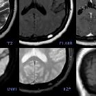
skull vault tumors in adults. Epidermoid cysts (a–c and d–f) and dermoid cyst (g–i). Case 1 (a–c): Extracranial soft-tissue mass (arrows) remodeling the outer table on NECT (a), with fluid T2-signal (b), and markedly restricted diffusion (b = 1000) (c). Case 2 (d–f): Transdiploic mass (arrowheads), predominantly intracranial, T1WI (d) and T2WI (e) heterogenous and intense diffusion restriction (f). Case 3 (g–i). Frontal skull lesion (dashed arrows) disrupting the inner table on CT (g, h); content is CT-hypodense and T1-hyperintense, corresponding to fat (i)
Epidermoid in der Kalotte
Siehe auch:
- Läsionen der Schädelkalotte
- fibröse Dysplasie der Kalotte
- Dermoid Schädelkalotte
- Epidermoid
- Epidermoidzyste vs Dermoidzyste
und weiter:


 Assoziationen und Differentialdiagnosen zu Epidermoid in der Kalotte: