Epidermoidzyste

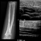
Nuchales
Atherom in der MRT: Oben T2 und DWi axial, unten FLAIR und T1 KM sagittal.

Epidermal
inclusion cyst • Epidermal inclusion cyst - Ganzer Fall bei Radiopaedia

Atherom der
Kopfhaut links hochparietal: In der Computertomografie reicht der Befund bis unmittelbar an die Hautoberfläche heran. Randständig betonte Verkalkungen. Knochenfenster links, im Weichteilfenster rechts oben, Hirnfenster rechts unten.

Breast
sebaceous cyst • Sebaceous cyst - MRI - Ganzer Fall bei Radiopaedia

Epidermal
inclusion cyst • Epidermal cysts - Ganzer Fall bei Radiopaedia

Epidermal
inclusion cyst • Epidermal inclusion cyst - ruptured - Ganzer Fall bei Radiopaedia

Epidermal
inclusion cyst • Epidermal inclusion cyst - neck - Ganzer Fall bei Radiopaedia

Epidermal
inclusion cyst • Leaked epidermal inclusion cyst - arm - Ganzer Fall bei Radiopaedia

Acoustic
enhancement • Epidermal inclusion cyst (gluteal region) - Ganzer Fall bei Radiopaedia

Epidermal
inclusion cyst • Epidermal cyst overlying the sacrum - Ganzer Fall bei Radiopaedia

Epidermal
inclusion cyst • Infrapatellar epidermal inclusion cyst - Ganzer Fall bei Radiopaedia

Epidermal
inclusion cyst • Frontal epidermal inclusion cyst - Ganzer Fall bei Radiopaedia

Epidermal
inclusion cyst • Intraosseous epidermoid cyst - probable - Ganzer Fall bei Radiopaedia

Epidermal
inclusion cyst • Sebaceous cyst - knee - Ganzer Fall bei Radiopaedia

Epidermal
inclusion cyst • Epidermal inclusion cyst: sole of foot - Ganzer Fall bei Radiopaedia

Epidermal
inclusion cyst • Epidermal inclusion cyst - parotid region - Ganzer Fall bei Radiopaedia

Epidermal
inclusion cyst • Epidermal inclusion cyst - Ganzer Fall bei Radiopaedia

Pseudotumoural
soft tissue lesions of the foot and ankle: a pictorial review. Epidermoid cyst (different patient from that in Fig. 7). Axial fat-suppressed T2-WI (a), coronal T1-WI (b) and coronal contrast-enhanced T1-WI (c). Unilocular, well-defined subcutaneous lesion (arrows) at the medial aspect of the ankle. The lesion has intermediate SI on T2-WI with some small areas of lower SI (probably due to keratinous debris), and is slightly hyperintense to muscle on T1-WI. A peripheral rim enhancement is seen (arrowhead), without internal enhancement

Epidermal
inclusion cyst • Epidermal inclusion cyst of the external auditory canal - Ganzer Fall bei Radiopaedia

Splenic cyst
• Splenic epidermoid cyst - Ganzer Fall bei Radiopaedia

Epidermoid
cyst • Epidermoid cyst - Ganzer Fall bei Radiopaedia

Epidermoid
cyst • Intrascrotal extratesticular epidermoid cyst - Ganzer Fall bei Radiopaedia

Epidermoid
cyst • Epidermoid cyst - Ganzer Fall bei Radiopaedia

Breast
sebaceous cyst • Sebaceous cyst - breast - Ganzer Fall bei Radiopaedia

Epidermal
inclusion cyst • Epidermal inclusion cyst - Ganzer Fall bei Radiopaedia

Breast
sebaceous cyst • Sebaceous cyst - breast - Ganzer Fall bei Radiopaedia

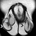
Intrakranielle
Epidermoidzyste der Pinealisregion rechts paramedian. Magentresonanztomographie DWI axial: Der Befund zeigt sich deutlich hyperintens.

Epidermal
inclusion cyst of the tongue - a rare entity. Axial T1 with contrast shows a well-defined lesion (1.5x1.0x1.2 cm) with mild peripheral enhancement and low signal intensity.
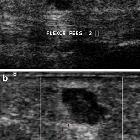
Pseudotumoural
soft tissue lesions of the foot and ankle: a pictorial review. Ultrasound of epidermoid cyst on the sole of the foot. The ultrasound images demonstrate a hypoechogenic polylobular lesion with posterior acoustic enhancement containing some scattered echogenic reflective material. The cyst is located in the subcutaneous fat superficial to the flexor tendon of the second digit

Extratesticular
epidermal inclusion cyst of the scrotum (ECR 2016 Case of the Day). Transverse T1-weighted image depicts homogeneous right extratesticular mass (arrow), isointense to the ispilateral testis (arrowhead).
Extratesticular
epidermal inclusion cyst of the scrotum (ECR 2016 Case of the Day). Coronal T2-weighted image shows right extratesticular mass (arrow), close to the ipsilateral spermatic cord (long arrow), mainly hyperintense, with signal similar to that of normal testes (arrowheads), surrounded by a low signal intensity capsule.

A rare case
of extratesticular scrotal epidermoid cyst. A non-vascular lesion measuring 15x12 mm was noted indenting the lateral aspect of the right epididymal head with a whorled appearance and central calcific focus giving it a "target" or "bulls eye" appearance.

Epidermal
cyst of the ischiorectal fossa. No rim enhancement is appreciable on axial post-contrast image.
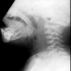
Toddler with
stridorLateral radiograph of the airway shows a mass at the base of the tongue.The diagnosis was epidermal inclusion cyst of tongue.

Intrakranielle
Epidermoidzyste der Pinealisregion rechts paramedian. Magentresonanztomographie T1w axial nach Kontrastmittel: Keine KM-Aufnahme


Epidermal
inclusion cyst • Sebaceous cyst - neck - Ganzer Fall bei Radiopaedia

Intrakranielle
Epidermoidzyste der Pinealisregion rechts paramedian. Magentresonanztomographie T2w sagittal: Der Befund zeigt sich nahezu liquorisointens. Aber siehe FLAIR und DWI.

Intrakranielle
Epidermoidzyste der Pinealisregion rechts paramedian. Magentresonanztomographie T2w axial: Der Befund zeigt sich nahezu liquorisointens. Aber siehe FLAIR und DWI.

Intrakranielle
Epidermoidzyste der Pinealisregion rechts paramedian. Magentresonanztomographie T1w coronar nach Kontrastmittel: Keine KM-Aufnahme

Epidermal
inclusion cyst • Epidermal inclusion cyst (forearm) - Ganzer Fall bei Radiopaedia

Epidermal
inclusion cyst • Epidermal inclusion cyst - supraorbital region - Ganzer Fall bei Radiopaedia

Epidermal
inclusion cyst • Epidermal inclusion cyst - Ganzer Fall bei Radiopaedia

Epidermal
cyst of the ischiorectal fossa. A well-circumscribed large cyst occupies the right ischiorectal fossa; the rectum and the vagina are contralaterally displaced.

Epidermal
cyst of the ischiorectal fossa. Axial image: the cyst is unilocular and delineate by thin hypointense rim.


Pseudotumoural
soft tissue lesions of the hand and wrist: a pictorial review. Subcutaneous epidermoid cyst with involvement of the adjacent bone of the terminal phalanx of the right fifth finger. a Plain radiograph showing a well-defined osteolytic defect at the radial side of the distal phalanx. Note cortical destruction of the radial cortex. b Coronal SE T1-WI. The lesion is of intermediate signal intensity (arrows) with some internal areas of relatively high signal. c Coronal TSE T2-WI. High signal intensity of the lesion (arrows). d Coronal FS SE T1-WI after intravenous injection of gadolinium contrast medium. There is only subtle peripheral enhancement (arrows) of the lesion
Epidermoid cysts are nonneoplastic inclusion cysts derived from ectoderm that are lined solely by squamous epithelium. These are discussed separately by anatomic location:
- epidermal inclusion cyst
- intracranial epidermoid cyst
- splenic epidermoid cyst
- spinal epidermoid cyst
- testicular epidermoid cyst
See also
Siehe auch:
- intrakranielle Epidermoidzyste
- Ganglion (Überbein)
- Neurofibrom
- Epidermoid in der Kalotte
- Epidermoidzyste Finger
- Dermatofibrosarcoma protuberans
- epidermale Inklusionszyste der Mamma
- testicular epidermoid cyst
- intraventricular epidermoid
- Atherom Galea
- Fasciitis nodularis
- Epidermoidzyste vs Dermoidzyste
- kutane Metastasen
- Malherbe-Tumor
- atheroma
- reifes zystisches Teratom
und weiter:
- Mega Cisterna magna
- Arachnoidalzyste
- Rathke Zyste
- Cholesteatom
- Epidermoidzyste
- ASP-Assoziation
- Pinealiszyste
- Ecchordosis physaliphora
- Dermoidzyste
- Tumor Kleinhirnbrückenwinkel
- Blake's-Pouch-Zyste
- Dandy-Walker continuum
- neuroglial cyst
- epidermale Inklusionszyste
- Akroosteolyse
- ependymal cyst
- Keimzelltumor
- erworbenes Cholesteatom
- Cholesteatom des äußeren Gehörgangs
- zystische Läsionen in der Hypophyse
- Neoplasien der Cauda equina
- intraossäre Epidermoidzyste
- Steatocystoma multiplex
- hypothalamic lesions
- Dermoid Schädelkalotte
- testicular epidermoid
- congenital cholesteatoma
- white epidermoid
- periurethral cystic lesions
- Epidermoidzyste im Kleinhirnbrückenwinkel
- Epidermoid
- diffusionsgewichtete Bildgebung
- zystische Läsionen der Sellaregion
- Epidermoidzyste der Kalotte
- mostly / purely cystic pituitary region masses
- mikrozystisches Meningeom
- diffusion MRI of an epidermoid tumor
- epidermoid cyst of the cauda equina
- Läsionen der Fingerspitze
- extratestikuläre intraskrotale Epidermoidzyste
- cystic extraaxial mass
- sutural epidermoid cyst
- Merkspruch Keimzelltumoren
- cutaneous epidermal cyst
- epidermale Inklusionszyste der Zunge
- Nasoalveoläre Zyste


 Assoziationen und Differentialdiagnosen zu epidermale Inklusionszyste:


epidermale
Inklusionszyste der Mamma