cystic teratoma

Reifes
zystisches Teratom des Ovars (Dermoidzyste): Große Anteile mit fett-isodensen Werten, am Rand weichteildichte und kalkdichte Anteile.

A diagnostic
approach to the mediastinal masses. Chest imaging shows well the highly heterogeneous contents of mediastinal teratomas. a Mature cystic teratoma in a 40-year-old man. Contrast-enhanced CT scan shows a heterogeneous anterior mediastinal mass with areas of fat (open arrow), calcification (arrow) and fluid attenuation (*). Posterior displacement of mediastinal structures is also be seen. b Photograph of the surgical specimen. c Contrast-enhanced CT scan of an asymptomatic 24-year-old woman demonstrates a well-defined uniloculated mass located in prevascular space which shows a cystic changes within (*). Non foci of calcification were identified. d The mass was surgically removed and pathological examination confirmed a benign teratoma

Mature cystic
ovarian teratoma • Mature cystic ovarian teratoma - floating balls sign - Ganzer Fall bei Radiopaedia

Mature cystic
ovarian teratoma • Mature cystic ovarian teratoma - Ganzer Fall bei Radiopaedia

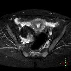
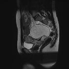
Ovarian
mature cystic teratoma: MR imaging findings. Axial T1-weighted image shows a left adnexal mass (arrow) with a fluid-fluid level and a floating solid element (long arrow), the latter corresponding to Rokitansky nodule on histology. Parts of the lesion appear hyperintense.

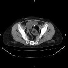
Ovarian
mature cystic teratoma: MR imaging findings. Axial T2-weighted image depicts heterogeneous left adnexal mass, mainly of high signal intensity. Normal right ovary (arrow) and uterus (long arrow).

Ovarian
mature cystic teratoma: MR imaging findings. Axial fat-suppressed T1-weighted image demonstrates saturation of the hyperintense T1 components (arrow, long arrow) of the lesion, findings compatible with the presence of fat.

Ovarian
mature cystic teratoma: MR imaging findings. ADC map derived from source image with b value of 1000 s/mm2 shows severely restricted diffusion of the fatty components (arrow, long arrow).
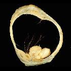
Mature cystic
ovarian teratoma • Mature cystic teratoma of the ovary (gross pathology) - Ganzer Fall bei Radiopaedia

Mature cystic
ovarian teratoma • Mature cystic ovarian teratoma - gross pathology - Ganzer Fall bei Radiopaedia
Mature cystic
ovarian teratoma • Ovarian dermoid cyst - Ganzer Fall bei Radiopaedia

Mature cystic
ovarian teratoma • Dermoid cyst of the ovary - Ganzer Fall bei Radiopaedia

Mature cystic
ovarian teratoma • Mature cystic ovarian teratoma - Ganzer Fall bei Radiopaedia

Mature cystic
ovarian teratoma • Ovarian mature cystic teratoma - Ganzer Fall bei Radiopaedia

Mature cystic
ovarian teratoma • Mature cystic ovarian teratoma - Ganzer Fall bei Radiopaedia

Mature cystic
ovarian teratoma • Mature (cystic) ovarian teratoma - Ganzer Fall bei Radiopaedia

Mature cystic
teratoma with torsion. A Lobulated lesion (red arrow) is seen on the right side in the pelvis. Enlarged left adnexa (blue arrow) is seen with twisted pedicle and midline displacement. Uterus is deviated to the right (green arrow).

Mature cystic
ovarian teratoma • Mature cystic ovarian teratoma - Ganzer Fall bei Radiopaedia

Mature cystic
ovarian teratoma • Mature cystic ovarian teratoma with Rokitansky nodule - Ganzer Fall bei Radiopaedia

Mature cystic
ovarian teratoma • Mature cystic ovarian teratoma - Ganzer Fall bei Radiopaedia

Mature cystic
ovarian teratoma • Ovarian dermoid cyst - pediatrics - Ganzer Fall bei Radiopaedia

Mature cystic
ovarian teratoma • Ovarian dermoid cyst - Ganzer Fall bei Radiopaedia

Mature cystic
ovarian teratoma • Right ovarian dermoid cyst in pregnancy (MRI) - Ganzer Fall bei Radiopaedia

Mature cystic
ovarian teratoma • Ovarian dermoid - Ganzer Fall bei Radiopaedia

Mature cystic
ovarian teratoma • Mature cystic ovarian teratoma - Ganzer Fall bei Radiopaedia

Mature cystic
ovarian teratoma • Ovarian dermoid cyst - Ganzer Fall bei Radiopaedia
Mature cystic
ovarian teratoma • Ovarian serous cystadenocarcinoma - Ganzer Fall bei Radiopaedia

Mature cystic
ovarian teratoma • Ovarian dermoid cyst - Ganzer Fall bei Radiopaedia

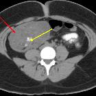
Mature cystic
teratoma with torsion. Lesion (red arrow) with fat and calcification (yellow arrow) within. Minimal free fluid is seen inferior to the lesion (orange arrow). Note right side displacement of uterus(green arrow) and enlarged left adnexa (blue arrow).

Mature cystic
teratoma with torsion. A lobulated lesion (red arrow) is seen in the right lower abdomen and pelvis with evidence of fat and calcification (tooth-like)(yellow arrow) within the lesion.

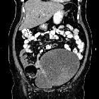
Typical CT
presentation of a mature cystic teratoma in a 53-years woman.. Basal CT shows an abdominal mass capsulated, inhomogeneous for the presence of materials with different density, included a calcification and fat, with fat-fluid level.

Mature cystic
ovarian teratoma • Ovarian mature cystic teratoma - Ganzer Fall bei Radiopaedia

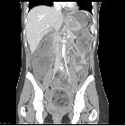
Typical CT
presentation of a mature cystic teratoma in a 53-years woman.. MPR in coronal and sagittal plane can help us and the surgeon to have a complete view of the tumour and the ralation between organs.

Typical CT
presentation of a mature cystic teratoma in a 53-years woman.. CECT shows a mild enhancement of the tumour"s walls.

Typical CT
presentation of a mature cystic teratoma in a 53-years woman.. A prone late scan is perfomed to demonstrate the fat-fluid level that is pathognomonic of a MCT. We also notice a Rokitansky nodule moving inside the MCT as the position changes.

Typical CT
presentation of a mature cystic teratoma in a 53-years woman.. An abdominal US shows a capsulated, inhomogeneous mass inside, occupying the four quadrants, of unknown origin.

Mature cystic
ovarian teratoma • Mature cystic teratoma - Ganzer Fall bei Radiopaedia

Mature cystic
ovarian teratoma • Mature cystic teratoma of ovary - Ganzer Fall bei Radiopaedia

Mature cystic
ovarian teratoma • Ovarian dermoid - Ganzer Fall bei Radiopaedia
Mature cystic
ovarian teratoma • Ruptured mature ovarian teratoma - Ganzer Fall bei Radiopaedia
Mature cystic
ovarian teratoma • Ovarian dermoid cyst - Ganzer Fall bei Radiopaedia

Mature cystic
ovarian teratoma • Ovarian dermoid cyst (MRI) - Ganzer Fall bei Radiopaedia

Mature cystic
ovarian teratoma • Ovarian dermoid - Ganzer Fall bei Radiopaedia

Mature cystic
ovarian teratoma • Ovarian torsion with dermoid cyst - Ganzer Fall bei Radiopaedia

Mature cystic
teratoma with torsion. A lobulated lesion (red arrow) is seen in the right lower abdomen and pelvis with evidence of fat and calcification (tooth-like)(yellow arrow) within the lesion.

Mature cystic
ovarian teratoma • Septate uterus with ovarian pathologies - Ganzer Fall bei Radiopaedia

Mature cystic
ovarian teratoma • Mature ovarian teratoma - Ganzer Fall bei Radiopaedia

Mature cystic
ovarian teratoma • Ovarian teratoma - Ganzer Fall bei Radiopaedia

Mature cystic
ovarian teratoma • Ovarian dermoid cyst with Rokitansky nodule - Ganzer Fall bei Radiopaedia
Mature cystic
ovarian teratoma • Mature cystic teratoma - Ganzer Fall bei Radiopaedia

Mature cystic
ovarian teratoma • Ovarian dermoid - Ganzer Fall bei Radiopaedia

Mature cystic
ovarian teratoma • Mature cystic ovarian teratoma - floating ball appearance - Ganzer Fall bei Radiopaedia

Reifes
zystisches Teratom des Ovars (Dermoidzyste): Große Anteile mit fett-isodensen Werten, am Rand weichteildichte und kalkdichte Anteile.

Mature cystic
teratoma with torsion. Lesion (red arrow) in the right lower abdomen and pelvis with fat and calcification (yellow arrow). Enlarged left adnexa (blue arrow) with twisted pedicle and midline displacement. Uterus deviated to the right(green arrow).


Mature cystic
teratoma with torsion. A Lobulated lesion (red arrow) is seen in the right lower abdomen with evidence of fat and calcification (tooth-like)(yellow arrow) within.
Mature cystic
teratoma with torsion. A Lobulated lesion (red arrow) is seen in the right lower abdomen with evidence of fat and calcification (tooth like)(yellow arrow) within.
Mature cystic
ovarian teratoma • Ovarian dermoid cyst complicated by torsion - Ganzer Fall bei Radiopaedia
Der Begriff reifes zystisches Teratom bezieht sich meist auf das reife zystische Teratom des Ovars. Andere Lokalisationen sind aber möglich.
Siehe auch:
- Teratom des Ovars
- eingeblutete Ovarialzyste
- Teratom
- Dermoidzyste
- Neoplasien des Ovars
- Lipoleiomyom des Uterus
- Rokitansky protuberance
- unreifes Teratom des Ovars
- muzinöses Zystadenom
- zystische Ovarialtumoren
- reifes zystisches Teratom des Ovars
- maligne Transformation bei Dermoidzyste des Ovars
- benigne Tumoren des Ovars
- fetthaltiger Dermoidtumor
- Keimzelltumor des Ovars
- Ruptur ovarielle Dermoidzyste
- ovarian serous
und weiter:


 Assoziationen und Differentialdiagnosen zu reifes zystisches Teratom:


