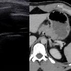
Extrapleural hematoma


Extrapleural hematomas are uncommon and usually seen in the context of rib fracture, subclavian venous catheter traumatic insertion, and blunt chest injury.
Pathology
Extrapleural hematomas result from the accumulation of blood in the extrapleural space where the overlying extrapleural fat is displaced centrally.
Etiology
- injury to intercostal arteries or veins in the setting of trauma
- traumatic insertion of a subclavian central venous catheter or intercostal catheter
- less commonly, from other vessels such as aortic rupture
Radiographic features
Plain radiograph
When located laterally, an extrapleural hematoma may cause the typical peripheral pleural opacity which has smooth borders with the pleura without acute angles. When anterior or posterior, the frontal X-ray will just show non-specific opacification.
CT
May show a focal extrapleural collection in the appropriate clinical context with an extrapleural fat sign. They may be biconvex or nonconvex, with the former being larger.
Treatment and prognosis
Biconvex extrapleural hematomas more often require surgical intervention, while non-convex hematomas are usually be managed conservatively.
Siehe auch:
- Pleuraempyem
- Rippenfraktur
- Hämatothorax
- tumorähnliche Läsionen der Pleura
- epipleurales Fett
- extrapleurales Fettzeichen
und weiter:

 Assoziationen und Differentialdiagnosen zu extrapleurales Hämatom:


