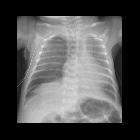
Intercostal catheter


Intercostal
catheter • Pneumothorax with displaced chest tube - Ganzer Fall bei Radiopaedia

Intercostal
catheter • Malpositioned intercostal catheter - Ganzer Fall bei Radiopaedia

Intercostal
catheter • Misplaced chest tube - Ganzer Fall bei Radiopaedia

Intercostal
catheter • Pneumocath in pulmonary artery and left atrium - Ganzer Fall bei Radiopaedia

Intercostal
catheter • Kinked intercostal catheter - Ganzer Fall bei Radiopaedia

Intercostal
catheter • Fractured left pleural catheter - Ganzer Fall bei Radiopaedia

Intercostal
catheter • Traumatic pneumothorax with malpositioned intercostal catheter - Ganzer Fall bei Radiopaedia

Intercostal
catheter • Displaced chest tube - Ganzer Fall bei Radiopaedia

Intercostal
catheter • Right lower lobe collapse - Ganzer Fall bei Radiopaedia

Intercostal
catheter • Chest tube in axilla - Ganzer Fall bei Radiopaedia
The intercostal catheter (ICC or chest tube) is a tube inserted into the pleural space to drain gas or fluid. It is mainly inserted to treat pneumothorax.
Indication
The indications are wide and can include :
- pneumothorax
- chest trauma
- pleural effusion
- hemothorax
- chylothorax
- bronchopleural fistula
- post-cardiothoracic surgery
- parapneumonic effusion or empyema
Classification
- ICC
- pleuracath
- pleural pigtail catheter
Site
ICCs are best placed in the triangle of safety, especially when put in urgently.
Placement
- consent
- patient position
- preferred: sitting at a 30-45° incline with the arm on side of the procedure abducted
- alternative: sitting upright and supported onto a table anteriorly or lateral decubitus
- identify triangle of safety, insertions are preferably oriented slightly superior to the border of the inferior rib to minimize the risk of damaging the neurovascular bundle
- sterile preparation and drape
- local anesthetic infiltration down to the level of parietal pleura
- 1-2 cm incision parallel to the rib
- blunt dissection using index finger or blunt forceps and breech parietal pleura: will be accompanied by a release of blood, fluid or air
- dilate insertion site with index finger
- insert drain with forceps directed towards the lung apex for pneumothorax or basally for hemothorax or fluid drainage
- drain size
- 10-14 Fr
- smaller bore drains are recommended to minimize discomfort
- larger bore drains are recommended for draining hemothoraces
- there should be minimal resistance during drain insertion and an appropriate length should be passed within the pleural cavity
- suture drain superficially and secure
- connect the drain to an underwater seal drain system
- confirmation: chest radiograph post procedure
- in certain drains, the absence of ‘swinging and bubbling’ in the underwater seal mechanism are concerning signs of dysfunction
Complications
- general complications
- malpositioning
- pain
- pneumothorax/tension pneumothorax
- infection
- device failure/malfunction
- complications arising from damage to local structures
- heart and great vessels
- lungs and tracheobronchial tree
- esophagus and stomach
- upper abdominal organs especially the liver and spleen
- intercostal nerves and vessels
Siehe auch:
und weiter:

 Assoziationen und Differentialdiagnosen zu Thoraxdrainage:
