Fehllage Thoraxdrainage

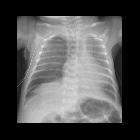
Newborn with
hydrops fetalis after bilateral chest tube placement. CXR AP shows body wall thickening. The tip of the left chest tube is in the left pleural space and there is a small left pleural effusion. The tip of the right chest tube is in the right subcutaneous tissues. There is a small right pleural effusionThe diagnosis was chest tube malfunction with the tip of the right chest tube in the subcutaneous tissues of the right chest wall in a patient with hydrops fetalis.
Premature
newborn after chest tube placementCXR AP shows diffuse ground glass opacity throughout the lungs and a large amount of air in the right pleural space causing mediastinal shift to the left while the right-sided chest tube courses through the subcutaneous tissues of the right chest wall and never enters the right pleural space.The diagnosis was persistent pneumothorax in a patient with respiratory distress syndrome due to the chest tube tip not being in the pleural space.

Young adult
with cystic fibrosis with continued shortness of breath after chest tube placementCXR AP (left) shows chronic interstitial fibrosis and scarring in the lungs, a left-sided chest tube, and a moderately-sized basilar left pleural air collection manifesting as a deep sulcus sign. Gross pathological specimen (right) shows the left chest tube entering the upper lobe of the left lung. The diagnosis was persistent pneumothorax in a patient with cystic fibrosis due to the chest tube tip not being in the pleural space.
Fehllage Thoraxdrainage
Siehe auch:


 Assoziationen und Differentialdiagnosen zu Fehllage Thoraxdrainage:

