fetal renal pelvic dilatation
Fetal pyelectasis refers to the prominence of the renal pelvis in utero that is a relatively common finding, which in the majority of cases resolves spontaneously.
Please refer to the article on fetal hydronephrosis for a continued discussion on this matter.
Terminology
Although there is an overlap of definition between pyelectasis and hydronephrosis, the former has been widely used instead of mild hydronephrosis given that the vast majority of the cases represent only a physiological incidental finding that resolves spontaneously, while the latter tends to be reserved for cases where a pathological obstruction is suspected.
Epidemiology
Fetal pyelectasis can be a relatively common finding on antenatal ultrasounds, often detected at the routine 2nd-trimester morphology scan. There is a recognized male predilection. The estimated prevalence is at ~2% of routine second trimester scans .
Pathology
Pyelectasis can result from a number of factors. In the majority of cases, it is physiological and resolves spontaneously. However, it may also herald the presence or evolution of renal tract pathology, such as:
- fetal PUJ obstruction
- fetal VUJ obstruction
- urethral obstruction, e.g. posterior urethral valves
- vesicoureteric reflux
- duplex kidney
Associations
- trisomy 21: isolated fetal renal pyelectasis is not considered to increase the risk of aneuploidies, however, in combination with other soft markers, the risk will increase (see antenatal features of Down syndrome)
Radiographic features
Ultrasound
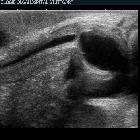
Fetal pyelectasis is assessed as an AP measurement of the renal pelves on an axial plane ultrasound image.
According to the Society of Fetal Urology (SFU) consensus, fetal pyelectasis is considered present if the anterior to posterior diameter of the renal pelvis measures:
- >4 mm up to 28 weeks
or - >7 mm at or after 28 weeks gestation
Note should also be made of any renal calyceal dilatation, ureteric dilatation, renal parenchymal appearance, bladder appearance and any unexplained oligohydramnios.
Fetal pyelectasis can also be affected by maternal hydration, i.e. physiological fetal pyelectasis .
Treatment and prognosis
The vast majority of cases (~96%) with mild pyelectasis in the second trimester resolve, either during pregnancy or in the early postpartum period. The risk of postnatal renal pathology is increased with:
- increasing degree of pelvic dilatation
- in utero progression
- bilateral involvement
Antenatally detected renal pelvic dilatation, especially in isolation, is considered a weak predictor of vesicoureteric reflux although postnatal sonographic evaluation is often recommended.
The finding of pyelectasis in the second trimester is typically monitored with a repeat prenatal scan in the third trimester, with further postnatal follow-up recommended in cases of persistent pyelectasis .
Postnatally, most cases with pyelectasis resolve spontaneously in the first year of life, and invasive procedures are not required.
Siehe auch:
- Down-Syndrom
- Doppelniere
- fetal hydronephrosis
- Urethralklappe
- Vesikoureteraler Reflux
- fetal PUJ obstruction
und weiter:


 Assoziationen und Differentialdiagnosen zu fetal pyelectasis: