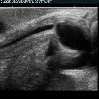
fetal hydronephrosis
Fetal hydronephrosis represents the abnormal dilatation of the fetal renal collecting system, with pelviureteric junction obstruction the most commonly encountered cause.
Please, refer to the article on fetal pyelectasis for a dedicated discussion on this relatively common and usually benign form of mild hydronephrosis.
Terminology
Although there is an overlap of definition between pyelectasis and hydronephrosis, the former has been widely used instead of mild hydronephrosis given that the great majority of the cases represent only a physiological incidental finding that resolves spontaneously, while the latter tends to be reserved for cases where a pathological obstruction is suspected.
Epidemiology
Depending on its definition, the estimated prevalence may range between 1-3% of pregnancies.
Pathology
Marked hydronephrosis (particularly in the 3 trimester) can result from a number of pathologies, which include:
- posterior urethral valves: in males
- congenital pelviureteric junction obstruction: the commonest cause of fetal hydronephrosis
- congenital vesicoureteric junction obstruction
- ureterocoele(s)
- urethral agenesis
- congenital megaloureter
- megacystis microcolon syndrome
- megacystis megaureter syndrome
- fetal vesicoureteric reflux
- prune-belly syndrome (rare)
Associations
- Down syndrome: more so with mild renal pelvic dilatation (low association)
Radiographic features
Antenatal ultrasound
In the second trimester, the severity of fetal hydronephrosis may be graded as .
- mild hydronephrosis: anteroposterior renal pelvic diameter measures ≥5mm (≥4mm at 16-20 weeks)
- moderate/severe hydronephrosis: anteroposterior renal pelvic diameter measures ≥7 mm or if there is associated calyceal dilatation
- persistent hydronephrosis: ≥10 mm in the 3 trimester
Ancillary sonographic features:
dilatation of the renal calyces
- dilated ureter if the obstruction is distal
- concurrent oligohydramnios if there is a bilateral obstruction (or unilateral obstruction with significantly impaired renal function in the other kidney)
Treatment and prognosis
Management will depend on the underlying pathology. The degree of renal pelvic dilatation correlates with the outcome. A renal pelvic anteroposterior diameter of 9 mm or more, and a pelvic-to-renal anteroposterior diameter ratio of 0.45 before 32 weeks of gestation and 0.52 thereafter are considered to be useful for the detection of a severe outcome postnatally .
Siehe auch:
- Down-Syndrom
- Ureterozele
- Oligohydramnion
- Vesikoureteraler Reflux
- fetal pyelectasis
- Urethralklappe
- urethral atresia
- kongenitaler Megaureter
- megacystis megaureter syndrome
- pelvi-ureteric junction obstruction
- mild renal pelvic dilatation
- megacystis micro-colon syndrome
und weiter:

 Assoziationen und Differentialdiagnosen zu fetal hydronephrosis:


