prune belly syndrome


Prune belly syndrome, also known as Eagle Barrett syndrome or triad syndrome, is a rare anomaly comprising a specific constellation of features. It consists of three major findings:
- gross pelvicalyceal and ureteric dilatation with renal dysplasia
- anterior abdominal wall underdevelopment (resulting in the "prune belly" appearance)
- bilateral undescended testes (cryptorchidism) in males
There is often an association with other respiratory, gastrointestinal, musculoskeletal, and cardiovascular anomalies.
Epidemiology
The estimated incidence is at ~1 in 35,000-50,000 live births. It occurs almost exclusively in males (>95% ) and is seen more frequently in twin pregnancies.
Associations
- aneuploidic syndromic associations
- other associations
Clinical presentation
The ureters are most dilated distally, with the renal pelvis demonstrating disproportionately little dilatation. The renal parenchyma is usually well maintained, and renal function is good. Stasis makes infections and stones the biggest problems.
Urinary tract abnormalities include:
- bilateral hydroureteronephrosis: often with extremely dilated, tortuous ureters
- varying degrees of renal dysplasia
- enlarged urinary bladder, often with urachal diverticulum
- vesicoureteral reflux is common
- poor bladder contractility
- dilated posterior urethra without urethral obstruction
Pathology
The condition is of unknown cause. One theory suggests that there is a mesenchymal insult to the fetus at ~6 weeks gestation resulting in deficient abdominal muscular development. A second theory suggests that the problem may be secondary to chronic intrauterine abdominal distention with subsequent pressure atrophy of abdominal muscles .
Spectrum of severity
Prune belly syndrome occurs with variable degrees of severity. In severe cases, renal dysplasia and oligohydramnios in utero result in pulmonary hypoplasia. These infants may be stillborn or die shortly after birth often due to respiratory complications.
Those with less severe renal disease may survive infancy, but may have recurrent urinary tract infection or progressive renal insufficiency. Some mild cases may have little or no loss of renal function and therefore a better prognosis.
Radiographic features
Plain radiograph
Bulging abdomen due to lack of abdominal wall muscles .
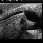
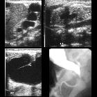
Ultrasound
Small kidneys with abnormal dilatation of calyces and ureters bilaterally. Large urinary bladder with thin wall and wide neck can be observed. There can be a patent urachus or urachal diverticulum. Ultrasound is used as a long term follow up .
MCUG
Important examination for urethral assessment. Risky for patient due to possibility of sepsis development. Two third of patients have vesicoureteric reflux .
Differential diagnosis
For antenatal hydronephrosis with hydroureter, consider:
- posterior urethral valves: may show a key hole sign and no evidence of cryptorchidism
- megacystis microcolon intestinal hypoperistalsis syndrome: tends to have polyhydramnios and more females affected
Siehe auch:
- Kryptorchismus
- Urethralklappe
- megacystis microcolon intenstinal hypoperistalsis syndrome
- abdominal wall aplasia
und weiter:


 Assoziationen und Differentialdiagnosen zu prune belly syndrome: