tetralogy of Fallot


Tetralogy of Fallot (TOF) is the most common cyanotic congenital heart condition with many cases presenting after the newborn period. It has been classically characterized by the combination of ventricular septal defect (VSD), right ventricular outflow tract obstruction (RVOTO), overriding aorta, and late right ventricular hypertrophy.
Epidemiology
Tetralogy of Fallot accounts for 5 to 10% of all congenital heart disease and has an estimated prevalence of 1 in 2000 births .
Associations
- cardiovascular associations
- right-sided aortic arch: seen in ~25% of cases
- pulmonary hypoplasia +/- atresia: particularly important in determining treatment , when there is pulmonary atresia it is sometimes termed pseudotruncus arteriosus
- atrial septal defect (ASD) or patent ductus arteriosus (PDA) (termed pentalogy of Fallot)
- coronary artery anomalies: seen in 3% of cases
- persistent left-sided superior vena cava
- extracardiovascular associations: may be present in ~15% of cases
Clinical presentation
The presentation relies on the degree of right ventricular outflow tract obstruction (RVOTO). Typically this is significant, resulting in cyanosis evident in the neonatal period, as a consequence of the right to left shunt across the ventricular septal defect. In cases where outflow obstruction is minimal, cyanosis may be inapparent (pink tetralogy) resulting in delayed presentation, even into adulthood, although this is rare.
Pathology
Tetralogy of Fallot is classically characterized by four features which are:
- maybe multiple in ~5% of cases
- infundibular stenosis, or
- hypoplastic pulmonary valve annulus, or
- bicuspid pulmonary valve, and/or
- hypoplasia of pulmonary artery
The right ventricular hypertrophy is a result of the ventricular septal defect and right ventricular outlet obstruction, both contributing to elevated resistance to right heart emptying .
Genetics
In ~15% of cases, it is associated with a deletion on chromosome 22q11 .
Radiographic features
Plain radiograph
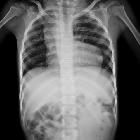
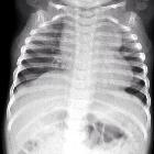
Chest radiographs may classically show a "boot-shaped" heart with an upturned cardiac apex due to right ventricular hypertrophy and concave pulmonary arterial segment. Most infants with tetralogy of Fallot, however, may not show this finding.
Pulmonary oligemia occurs due to decreased pulmonary arterial flow. A right-sided aortic arch is seen in 25%.
Echocardiography
Echocardiography allows direct visualization of the abnormal anatomy and remains the primary modality for the diagnosis of tetralogy of Fallot. It has limitations on assessing associated extracardiac anomalies (e.g. peripheral pulmonary stenosis and atresia). Salient features which may be assessed during a standard sequence of transthoracic echocardiographic views include ;
- parasternal long-axis view
- left and right ventricular size/function
- degree of aortic override
- override of the aortic root over the ventricular septal defect should be less than half of the aortic diameter
- analysis of the ventricular septal defect
- magnitude and direction of shunting across the VSD with spectral and color flow Doppler
- confirmation of aorto-mitral continuity
- absence of the fibrous continuity between the aortic and mitral valves is inconsistent with tetralogy of fallot, and may be suggestive of double outlet right ventricle (DORV)
- parasternal short-axis view
- location of the ventricular septal defect
- anatomy of the right ventricular outflow tract
- dilation of the RVOT may be observed
- dynamic obstruction of the RVOT may be noted, resulting from incursion of the infundibulum into the outflow tract
- continuous-wave Doppler through the RVOT may demonstrate a late-peaking, high-velocity envelope is consistent with dynamic obstruction
- right ventricular systolic pressure (RVSP) may also be quantified using the velocity of the tricuspid regurgitant jet if visible
- pulmonic valve
- leaflet number and degree of mobility
- dimensions of the annulus
- spectral Doppler interrogation for the presence and grading of associated pulmonic regurgitation or stenosis
- presence or absence of associated coronary artery anomalies
- apical 4 chamber view
- assess right and left ventricular size and function
- right ventricular hypertrophy may be assessed by measurement of the free wall of the right ventricle
- assess right and left ventricular size and function
CT
CT is useful in demonstrating the complex cardiovascular morphology of tetralogy of Fallot, especially the anatomy of the pulmonary and coronary arteries as well as identification of major aortopulmonary collateral arteries (MAPCAs). CT can be used to evaluate post-surgical changes (e.g. patency of palliative shunts) and complications.
MRI
MRI has the great advantage of providing both exquisite anatomical detail and functional information without ionizing radiation. A detailed assessment of the pulmonary artery is of particular importance because the repair of the cardiac defects without addressing pulmonary artery hypoplasia or stenosis has a poor outcome .
The main pulmonary artery or right pulmonary artery diameter should be compared to that of the ascending aorta. A ratio of <0.3 usually signifies that primary repair would be unsuccessful, and a bridging shunt operation may be of benefit .
Assessment of coronary artery origin is also essential to surgical planning.
Treatment and prognosis
Approximately 90% of untreated tetralogy of Fallot patients succumb by the age of 10 years . Over the years many surgical approaches were performed until the current primary repair was developed. Shunts are nowadays only performed as a palliative procedure in inoperable cases or to bridge patients until the repair can be carried out, typically in the setting of pulmonary arterial hypoplasia .
Shunt operations included :
- shunts: designed to reduce cyanosis
- Pott shunt
- Waterston shunt
- Blalock-Taussig shunt: still performed in selected cases
Primary repair (total correction (a.k.a. repair) of tetralogy of Fallot) is now the preferred treatment and is usually performed at the time of diagnosis.
Common post-surgical complications include :
- conduction abnormalities
- right bundle branch block (RBBB): 80-90% of cases
- bifascicular block: 15% of cases
- premature ventricular contractions (PVC): ~50% of cases
- sustained ventricular tachycardias (VT): ~5% of cases
- atrial arrhythmias: common
- valvular dysfunction
- tricuspid regurgitation
- pulmonary regurgitation
Prognosis is largely dependent on how soon the defect is diagnosed and corrected, with the best outcome seen in patients repaired before the age of 5 . Overall there is a 90-95% survival rate at 10 years of age, however, residual right ventricular dysfunction is common. Up to 10% of patients require reoperation within 20 years .
History and etymology
It is named after Étienne-Louis Arthur Fallot (1850-1911) , a French physician, who described the tetralogy of defects in 1888 .
Differential diagnosis
Findings on a chest radiograph are commonly non-specific, and other cyanotic congenital heart diseases should be considered.
Other conditions involving an outflow tract overriding a VSD are :
- pulmonary atresia with ventricular septal defect: considered an extreme form of tetralogy of Fallot, the pulmonary atresia is obstructed with anterograde flow. The ductus arteriosus is tortuous with retrograde flow in the pulmonary arteries. Surgery and prognosis is related to the anatomy of the pulmonary arteries
- type 1 features a simple diaphragm at the pulmonary annulus
- type 2 has no pulmonary trunk but the pulmonary arteries are preserved
- type 3 is like type 2 but also with atretic pulmonary arteries
- type 4 does not feature any pulmonary arteries but a systemic-pulmonary vascularization by major aortopulmonary collateral arteries (MAPCAs)
- absent pulmonary valve syndrome:
- can also be an associated feature with tetralogy of Fallot
- aortic root is overriding but not enlarged. PA trunk is, conversely, very large with to-and-fro flow due to both stenosis and insufficiency; ductus arteriosus is classically absent or atretic.
- common arterial trunk: pulmonary is arising from the overriding vessel, the common valve often regurgitates.
- double outlet right ventricle: mimics transposition of the great vessels, combined with a ventricular septal defect
Siehe auch:
und weiter:
- Ektasie Aorta ascendens
- Persistierender Ductus arteriosus
- Herzfehler
- Kardiomegalie
- Ebstein anomaly
- rechts descendierende Aorta
- Varianten der Herzanatomie
- antenatal features of Down syndrome
- Embryopathia rubeolosa
- CXR approach to congenital heart disease
- conotruncal cardiac anomalies
- Mikrodeletionsyndrom 22q11
- chest x-ray appeoach to congenital heart disease
- Pulmonalatresie
- tetralogy of fallot (mnemonic)
- congenital heart disease - chest x-ray approach
- Ventrikelseptumdefekt
- Thrombozytopenie-Radiusaplasie-Syndrom
- pentalogy of Fallot
- MRT-Angiographie
- Zyanotischer Herzfehler
- Cantrell’sche Pentalogie
- isolated unilateral absence pulmonary artery (IUAPA)
- Agenesie des Perikards
- pott shunt
- tetralogy of Fallot and tracheoesophageal fistula
- Herzektopie
- kongenitale Pulmonalstenose
- unusual communication between bilateral superior venae cavae in a baby with tetralogy of Fallot
- Blalock Taussig procedure
- Blalock Taussig shunt
- tetralogy of Fallot with absent pulmonary valve

 Assoziationen und Differentialdiagnosen zu Fallot'sche Tetralogie:

