22q11.2 deletion syndrome

22q11.2 deletion syndrome, also known as the DiGeorge syndrome or velocardiofacial syndrome, is a syndrome where a small portion of the chromosome 22 is lost and results in a variable but a recognisable pattern of physical and behavioral features.
Epidemiology
The estimated incidence is at ~ 1 in 4000-6000 live pregnancies .
Clinical presentation
CATCH 22 is the mnemonic to remember the chromosome and all the abnormalities.
- cleft lip +/- palate
- congenital heart disease (particularly conotruncal anomalies): often a major part of this syndrome
- characteristic facies
- elongated face
- short philtrum
- facial asymmetry
- prominent nose
- hypernasal speech
- learning disabilities
- decreased immunity
- malformation of third and fourth pharyngeal pouches that result in the defective development of the parathyroid and thymus which can, in turn, lead to
- hypoparathyroidism
- hypocalcemia
Pathology
Genetics
There is a near-universal association with a deletion within chromosome 22q11.2. The majority of cases have de novo mutations. 22q11 deletions are associated with some types of conotruncal cardiac defects as well as conotruncal anomaly face syndrome .
Associations
Radiographic features
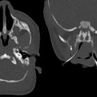
Temporal bone malformations are common: :
- lateral semicircular canal dysplasia with small or absent bony island, forming a wide or single cavity with the vestibule (42% and 26%, respectively)
- cochlear incomplete partition type II (32%)
- middle ear ossicles
- dense stapes superstructure (39%)
- malleus, incus, or other stapes abnormalities (7%)
- carotid canal dehiscence (10%)
Brain and cerebrovascular malformations are common :
- persistent cavum septi pellucidi and/or cavum vergae (19-33%)
- aberrant cortical veins (25%)
- polymicrogyria or cortical dysplasia (17%)
- white matter hyperintensities (10%)
- hypoplastic internal carotid artery (8%)
- brain volume loss, most pronounced in the cerebellum (see hypoplastic cerebellum)
Cardovascular, particularly conotruncal, defects are usually the first imaging abnormality noted in these patients :
- cardiac anomalies
- tetralogy of Fallot and variants (35%)
- interrupted aortic arch (type B) (19%)
- truncus arteriosus (9%)
- ventricular septal defect (16%)
- atrial septal defect (2%)
- transposition of the great arteries (2%)
- vascular anomalies
- right aortic arch (35%)
- vascular ring (5%)
- aberrant origin of subclavian artery (16%)
- mirror image brachiocephalic vessel branching (12%)
- left superior vena cava (9%)
History and etymology
First described in 1968 by Angelo DiGeorge (1921-2009), an American physician.
Siehe auch:
- Choanalatresie
- Fallot'sche Tetralogie
- Mondini-Dysplasie
- conotruncal cardiac anomalies
- langes Philtrum
- Atresie der Aorta
- Truncus arteriosus communis
- cleft lip - / + palate
und weiter:


 Assoziationen und Differentialdiagnosen zu Mikrodeletionsyndrom 22q11: