Fingerluxation


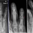
Joint
dislocation in the DIP of the third finger before (left images) and after (right images) reduction. Note that the dislocation can hardly be seen in the ap-projection (second image from left).


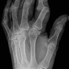
Joint
dislocation in the PIP of D4 and D5 before and after reduction. Note the little fragment (arrow) and the preexistent degenerative changes.


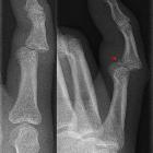
Teenager who
has pain in his fifth digit after it was hit by a football. AP (left), oblique (middle) and lateral (right) radiographs of the fifth digit show a complete dislocation of the fifth middle phalanx from the fifth proximal phalanx.The diagnosis was fracture dislocation of the fifth proximal interphalyngeal joint of the hand.


 Assoziationen und Differentialdiagnosen zu Fingerluxation:
