interphalangeal joint dislocation


Interphalangeal joint dislocations are common upper extremity dislocations. Although considered minor injuries by many, they can result in significant disability.
Pathology
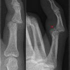
The typical mechanism is a hyperextension injury. The proximal interphalangeal joints are the most commonly involved and in the vast majority of cases, the dislocation is dorsal .
The proximal interphalangeal joints are mobile and stability is largely due to ligamentous support: collateral ligaments, volar plate, capsule, and the central slip of the extensor tendon . The volar plate, which is confluent with the periosteum of the phalanges, is key in maintaining joint stability and prevents hyperextension. In the majority of dorsal dislocations, it is damaged and may be associated with a small avulsion fracture . Collateral ligaments most commonly tear in the mid-substance, but can on occasion also result in an avulsion fracture .
Isolated dislocations of the distal interphalangeal joints are rare and usually are associated with avulsion fractures of the terminal extensor tendon or flexor digitorum profundus which insert into the base of the distal phalanges .
Radiographic features
Plain films are in almost all cases sufficient for the diagnosis. Ultrasound and/or MRI are reserved for assessment of ligamentous and tendinous structures in selected cases.
Plain radiograph
The diagnosis is usually self-evident provided adequate views are obtained and so long as the joint has not been reduced prior to imaging. Volar plate avulsion is often co-existant and should be specifically looked for in cases of dislocation.
Treatment and prognosis
Dislocations need to be reduced, preferably under a digital block . Most dorsal dislocations can be treated conservatively, even if they are associated with a small avulsion fracture (<25% of articular surface), provided they appear stable post reduction . Buddy splinting or an extension-block splint usually suffices .
In the rare instance of a volar dislocation, the central extensor slip is assumed to be avulsed. Immobilization in extension either with splinting or with K-wires is required for adequate healing .
If avulsion fractures are large (>25% of the articular surface) then they usually require internal fixation .
Practical points
In addition to stating that a dislocation is present a number of features should be sought and commented upon :
- dislocation: direction is predictive of the ligamentous structures likely to be damaged
- dorsal: most common, and associated with volar plate damage
- lateral/medial: collateral ligaments
- volar: uncommon, and associated with central extensor slip damage
- associated injuries
- fractures
- avulsion fracture: comment on % of articular surface involved
- open injury (gas in the soft tissues, usually clinically obvious)
- foreign bodies
- fractures
- post reduction
- when separation of the dorsal middle phalanges bases from the head of the proximal phalanx can be seen on the post-reduction lateral radiography it can indicate instability, this is known as a "V sign"
Siehe auch:


 Assoziationen und Differentialdiagnosen zu interphalangeal joint dislocation: