fractures of the atlas
Teenager with
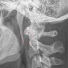
neck pain after hitting a tree with an all terrain vehicleAxial CT without contrast of the cervical spine shows a lucency through the anterior arch of the C1 vertebral body.The diagnosis was a non-displaced fracture of the anterior arch of C1.

Praktisch
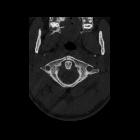
unverschobene, mehrfache Fraktur des Atlas im Sinne einer Jefferson-Fraktur bei einem 84 Jahre alten Mann nach Sturz auf den Kopf. Man erkennt in den beiden axialen Schichten der Computertomographie die Frakturlinien ventral und beidseits lateral. Keine Verlagerung des Dens.

Teenager who
has neck pain after a diving accidentAxial CT without contrast of the cervical spine shows lucencies through the left anterior and posterior arches of the C1 vertebral body with minimal displacement of the fracture fragments.The diagnosis was a minimally displaced fracture of the anterior and posterior arches of C1, a Jefferson fracture.

Teenager with
neck pain who hit his head on the ground while wrestling with a friendAxial CT without contrast of the cervical spine (upper left) shows lucencies through the left anterior and posterior arches of the C1 vertebral body with displacement of the fracture fragments. Coronal 2-D reconstruction (lower left) and the 3-D reconstruction (right) which simulates an open-mouth odontoid radiograph shows the lateral masses of the C1 vertebral body are now wider than the lateral masses of the C2 vertebral body.The diagnosis was displaced fracture of the anterior and posterior arches of C1, a Jefferson fracture.
Atlasfrakturen
fractures of the atlas
Siehe auch:
- Atlas Normvarianten
- Jefferson-Fraktur
- Halswirbelsäulenverletzung
- Klassifikation Atlasfrakturen nach Gehweiler
und weiter:


 Assoziationen und Differentialdiagnosen zu Atlasfrakturen: