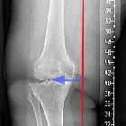
Genu valgum

Genu valgum (layperson term: knock-knee) denotes the valgus deformity of the knee, where the lower leg is bending outwards in relation to the axis of the femur.
Pathology
Etiology
Systemic conditions (e.g vitamin D deficiency) most commonly result in bilateral, whilst focal lesions (e.g. physeal trauma) in unilateral presentations .
Common causes:
- congenital
- physiologic developmental (between the age of 2 to 6-7 years)
- rickets (vitamin D deficiency or refractory, caused by hypophosphatemia)
- osteochondrodysplasia
- multiple epiphyseal dysplasia
- osteogenesis imperfecta
- iliotibial band contracture
- arthritis
- prior trauma, infection, tumor: usually unilateral
Radiographic appearance
Plain radiograph
The valgus deformity can be quantified with the hip-knee-ankle angle (HKA), which measures the angle between the mechanical axis of the femur and the center of the ankle joint on AP, full-length, weight-bearing radiographs. The first line is drawn between the center of the femoral head and the femoral intercondylar point, while the second line runs from the tibial interspinous point to the tibial mid-plafond point. The angle between these lines determines the hip-knee-ankle angle. The normal range is varying due to the physiologic varus angle in infants, and physiologic valgus configuration in young children, but is generally 1-1.5° in healthy adults .
Treatment and prognosis
Physiologic genu valgum is a self-correcting condition not necessitating treatment. For bilateral genu valgum caused by systemic disease, the treatment of the underlying condition is paramount. Unilateral genu valgum may necessitate surgical management (e.g. hemiepiphysiodesis, physeal tethering or distal femoral varus osteotomy) .
Siehe auch:
und weiter:

 Assoziationen und Differentialdiagnosen zu Genu valgum:


