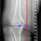
Genu varum


Genu varum (bow legs) denotes the varus angular deformity of the knee joint, where the leg is bowing outwards at the knee, while the lower leg is angled medially.
Pathology
Genu varum is physiologic in neonates and infants and reaches its peak between 6 to 12 months. During normal growth the tibiofemoral angle reaches zero between 18 to 24 months, after which it turns into a physiologic genu valgus, finally reaching the adult configuration by the age of 6 to 7 years. Genu varum after the age of 2 is considered to be abnormal .
Etiology
Common causes:
- physiologic (infants and neonates)
- rickets (vitamin D deficiency or refractory, caused by hypophosphatemia)
- bone dysplasia (e.g. achondroplasia)
- asymmetric arrested growth of the medial distal femur and proximal tibia (e.g. osteomyelitis, fracture, tumor)
- Blount disease (tibia vara)
- osteoarthritis
- prior trauma
Radiographic features
Plain radiograph
Proper radiographic assessment of genu varum necessitates standing AP and LL radiographs which should include the hip, knee and ankle. In pediatric patients, the growth plates should be evaluated carefully. In physiological genu varum, the bones demonstrate normal structure whilst e.g. rickets will show thickened physes, enlarged epiphyses, coarse bone trabeculae, and decreased cortical bone density .
The varus deformity can be quantified with the hip-knee-ankle angle (HKA), which measures the angle between the mechanical axis of the femur and the center of the ankle joint on AP, full-length, weight-bearing radiographs. The first line is drawn between the center of the femoral head and the femoral intercondylar point, while the second line runs from the tibial interspinous point to the tibial mid-plafond point. The angle between these lines determines the HKA. The normal range is varying due to the physiologic varus angle in infants but is generally 1-1.5° in healthy adults .
Treatment and prognosis
The management of genu varum primarily focuses on the treatment of the underlying condition (e.g. vitamin D supplementation in rickets). if conservative therapy (e.g. bracing, splints) fails, surgical management (valgus osteotomy of the tibia) can be considered .
Siehe auch:
und weiter:

 Assoziationen und Differentialdiagnosen zu Genu varum:

