achondroplasia


 nicht verwechseln mit: Achondrogenesie
Achondroplasia is a congenital genetic disorder resulting in rhizomelic dwarfism and is the most common skeletal dysplasia. It has numerous distinctive radiographic features.
Epidemiology
It occurs due to sporadic mutations in the majority of cases but can be inherited as an autosomal dominant condition. Homozygous achondroplasia is lethal.
There is a prevalence of approximately 1 in 25,000-50,000 births with males affected more frequently than females .
Clinical presentation
Achondroplasia is the most common cause of short-limb dwarfism. Patients are of normal intelligence with normal motor function. However, they may have specific neurologic deficits.
Pathology
The disease results from a mutation in the fibroblast growth factor gene 3 (FGFR3) located on chromosome 4p16.3 which causes abnormal cartilage formation. These are cell surface receptors comprised of an extracellular domain with three immunoglobulin-like regions, a transmembrane domain and an intracellular tyrosine kinase .
The mutation to the FGFR3 gene in achondroplasia is a gain of function mutation with constitutive activation of an inhibitory signal . All bones that form by endochondral ossification are affected. Bones that form by membranous ossification are not affected, thus allowing the skull vault to develop normally.
Associations
- SADDAN syndrome: severe achondroplasia with developmental delay and acanthosis nigricans
Radiographic features
Almost all the bones of the skeleton are affected, and hence all parts of the body have bony changes with secondary soft tissue changes. Antenatally it is difficult to diagnose achondroplastic features until the 3 trimester .
Antenatal ultrasound
Antenatally detectable sonographic features include:
- short femur length measurement: often well below the 5th centile
- the femur length (FL) to biparietal diameter (BPD) is taken as a useful measurement
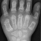
- trident hand : 2nd, 3rd and 4th fingers appearing separated and similar in length
- separation of 1st, 2nd, 3rd and 4th fingers
- protruding forehead: frontal bossing
- depressed nasal bridge
Plain radiograph / CT / MRI
Features on radiographs, CT, and MRI are similar and discussed together here.
Cranial
- relatively large cranial vault with small skull base
- frontal bossing with the depressed nasal bridge (midfacial retrusion)
- narrowed foramen magnum
- cervico-medullary kink
- relative elevation of the brainstem resulting in a large suprasellar cistern and vertically-oriented straight sinus
- communicating hydrocephalus (due to venous obstruction at sigmoid sinus)
- large anterior fontanelle in infancy; may persist to 5 or 6 years of age
Also, see the achondroplastic base of skull abnormalities for further discussion.
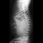
Spinal
- posterior vertebral scalloping
- progressive decrease in the interpedicular distance in the lumbar spine
- gibbus: thoracolumbar kyphosis with bullet-shaped/hypoplastic vertebra (not to be confused with Hurler syndrome)
- short pedicle canal stenosis
- laminar thickening
- widening of intervertebral discs
- an increased angle between the sacrum and lumbar spine
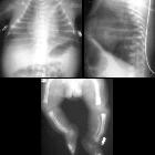
Chest
- anterior flaring of the ribs
- anteroposterior narrowing of the ribs
Pelvis and hips
- horizontal acetabular roof (decreased acetabular angle)
- small squared (tombstone or mickey mouse ear) iliac wings
- small trident pelvis
- champagne glass type pelvic inlet
- short sacroiliac notches
Limbs
- metaphyseal flaring: can give a trumpet bone type appearance
- the femora and humeri are particularly shortened (rhizomelic shortening)
- long fibula: the fibular head is at the level of the tibial plateau (case 3)
- bowing to mesial segment of legs
- the limbs may also appear thickened but are in fact normal in absolute terms; thickening is perceived due to reduced length
- trident hand
- chevron sign
- the metacarpal and metatarsal bones, and in some cases the proximal phalanges, are short and of similar length
Treatment and prognosis
There is often a danger of cervical cord compression due to narrowing of the foramen magnum.
Treatment varies and is usually orthopedic, particularly to correct kyphoscolioses, as well as neurosurgical, to decompress the foramen magnum or shunt hydrocephalus .
Overall prognosis is good, with near-normal life expectancy in heterozygous individuals. When homozygous, the condition is fatal due to respiratory failure .
History and etymology
Achondroplasia literally means "without cartilage formation", although the pathology is impaired endochondral ossification (see Pathology above).
Differential diagnosis
The differential diagnosis is that of other less common skeletal dysplasias, including :
- achondrogenesis
- campomelic dysplasia
- thanatophoric dysplasia
- chondroectodermal dysplasia (Ellis-van Creveld syndrome)
See also
- causes of limb bowing: general differential for (lower) limb bowing
- rhizomelic dwarfism: general differential for rhizomelic limb shortening
Siehe auch:
- Rhizomelie
- Skelettdysplasie
- Thanatophore Dysplasie
- Ellis-van-Creveld-Syndrom
- camptomelic dysplasia
- chondroectodermal dysplasia
- Verbiegung der langen Röhrenknochen
- short limb skeletal dysplasias
- Achondrogenesie
- Knorpel-Haar-Hypoplasie
- Foramen magnum Stenose bei Achondroplasie
- Hypochondroplasie
- Achondroplasie Wirbelsäule
und weiter:
- trident hand
- Platyspondylie
- Basiläre Impression
- Aseptische Wirbelkörpernekrose
- Vertebra plana
- anterior vertebral body beaking
- frontal bossing
- Biegungsbruch
- scalloping Wirbelkörper
- short limb skeletal dysplasia
- Makrozephalie
- Gibbus
- Auftreibung Metaphysen
- low lying patella
- intraspinale zystische Läsionen
- Tibiaverbiegung bei Kindern
- inanimate object inspired signs
- Madelung-Deformität
- congenital syndromes associated with enlarged ventricles
- paediatric curriculum
- basilar invagination (mnemonic)
- Pyle-Syndrom
- narrow fetal thorax
- Pseudoachondroplasie
- Erlenmeyer flask deformity of the femur
- Acetabulumwinkel
- bilateral megalencephaly
- Mikromelie
- SADDAN syndrome
- Megaloenzephalie
- tentorial angle
- zapfenförmige Epiphyse
- vertically-oriented straight sinus
- J-förmige Sella turcica
- verminderter interpediculärer Abstand
- Mickey Maus Beckenschaufeln
- kugelförmige oder ovoide Wirbelkörper
- champagne glass pelvis


 Assoziationen und Differentialdiagnosen zu Achondroplasie: