congenital syndromes associated with enlarged ventricles

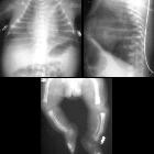
Newborn with
ventriculomegaly on prenatal ultrasound. Axial (above left) and coronal (above right) T1 MRI without contrast of the brain show dilation of the lateral and third ventricles. Sagittal T2 MRI without contrast of the brain (below) shows narrowing at the aqueduct of Silvius with a decreased amount of CSF within it. The fourth ventricle is normal in size.The diagnosis was aqueductal stenosis.
Congenital ventriculomegaly can have a large number of syndromic associations.
Common
- acrocephalosyndactylies
- acrocephalopolysyndactylies
- achondroplasia
- fetal alcohol syndrome
- lissencephaly
- osteopetrosis
- Sotos syndrome
- X-linked hydrocephalus syndrome
Uncommon
- Fryns syndrome
- Goldenhar syndrome
- hydrolethalus
- metachromatic leukodystrophy
- Miller-Dieker syndrome : also type of lissencephaly
- mucopolysaccharidoses (IH and VI)
- multiple pterygium syndrome
- Neu-Laxova syndrome
- skeletal dysplasias
- VACTERL-H association
- Walker-Warburg syndrome
Siehe auch:
- Achondroplasie
- Osteopetrose
- Lissenzephalie
- Apert-Syndrom
- Chondrodysplasia punctata
- Skelettdysplasie
- Thanatophore Dysplasie
- Mukopolysaccharidose
- Crouzon-Syndrom
- Walker-Warburg-Syndrom
- acrocephalosyndactyly
- camptomelic dysplasia
- Goldenhar-Gorlin-Syndrom
- Fetales Alkoholsyndrom
- Fryns-Syndrom
- Metachromatische Leukodystrophie
- Sotos-Syndrom
- acrocephalopolysyndactyly
- hydrolethalus
- Neu-Laxova-Syndrom
- Miller-Dieker Syndrom
- VACTERL-H
- multiple pterygium syndrome
und weiter:


 Assoziationen und Differentialdiagnosen zu congenital syndromes associated with enlarged ventricles: