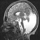
enlarged fetal ventricles


Fetal ventriculomegaly refers to the presence of dilated cerebral ventricles in utero.
Important in itself, it is also associated with other CNS anomalies.
Epidemiology
Using the current sonographic cut-off criteria (see radiographic features below), the estimated prevalence may be at ~0.9% of all pregnancies . There may be a slightly increased male predilection.
Pathology
Development of lateral ventricles
- first trimester
- the choroid plexus regularly fills the entire lateral ventricle, bilaterally
- second trimester
- the choroid plexus begins to recede posteriorly but remains in close contact with the medial and lateral walls of the bodies and atria of the ventricles
- likewise, the lateral cerebral ventricle is large relative to the cerebral hemispheric width
Causes
See the article: fetal ventriculomegaly (differential)
Associations
While many fetuses with mild ventriculomegaly have a normal outcome, there are also a large number of congenital syndromes associated with enlarged ventricles.
Radiographic features
Antenatal ultrasound
Ultrasound is the screening modality of choice for initial evaluation .
The measurement should be in the true axial plane at the atria of the lateral ventricle and glomus of the choroid plexus. The ventricle is measured from inner margin of the medial ventricular wall to inner margin of the lateral wall.
Fetal ventriculomegaly is defined as:
- >10 mm across the atria of the posterior or anterior horn of lateral ventricles at any point in the gestation
- alternatively, a separation of more than 3 mm of the choroid plexus from the medial wall of the lateral ventricle may be used
The severity of ventriculomegaly can be further classified as :
- mild/borderline fetal ventriculomegaly: lateral ventricular diameter between 10-12 mm
- moderate fetal ventriculomegaly: 12.1-15 mm
- severe fetal ventriculomegaly (also sometimes classified as fetal hydrocephalus): lateral ventricular diameter >15 mm
When ventriculomegaly is pronounced, the choroid plexus will no longer lie in an almost parallel fashion against the lateral ventricular wall. Tethered at the foramen of Monro the free hanging choroid will "hang down" and appear to "dangle" within the dilated ventricle. This appearance is often termed the dangling choroid sign. The ventricle to cerebral hemisphere ratio would also increase as a result.
Fetal brain MRI
MRI may be useful for evaluation of additional anomalies.
(more content required)
Significance when detected on ultrasound
Even when noted without an associated structural anomaly, mild fetal ventriculomegaly is often considered a soft antenatal marker for underlying chromosomal abnormalities. Therefore, a careful search for other sonographic abnormalities is recommended.
Careful ultrasound evaluation of the posterior fossa is also critical to look for a potential cause of obstructive hydrocephalus.
Borderline to mild prenatally detected ventriculomegaly, without additional abnormalities or an abnormal karyotype, the majority of children have been to reported to have a normal development .
Treatment and prognosis
The prognosis, as well as management, largely depend on the etiology and on the presence of associated abnormalities.
Differential diagnosis
- pseudo-hydrocephalus: if the ventricle appears enlarged, but there is no dangling choroid, the cerebrum may just be hypoechoic
- investigate closely, try different angles to find both hyperechoic lines of the lateral ventricle
See also
- fetal hydrocephalus: lateral ventricular diameter >15 mm
- fetal ventriculomegaly (differential)
Siehe auch:
- Arnold-Chiari-Malformation Typ 2
- Aquäduktstenose
- fetal toxoplasmosis
- congenital syndromes associated with enlarged ventricles
- Dysgenesie des Corpus callosum
- Dandy Walker continuum
- connatale Zytomegalie
- kongenitaler Hydrozephalus
- soft antenatal marker
und weiter:
- Spina bifida
- Meckel-Syndrom
- Arthrogryposis multiplex congenita
- antenatal features of Down syndrome
- Myeloschisis
- sonographic values in obstetrics and gynaecology
- Jarcho-Levin-Syndrom
- dysencephalia spanchnocystica
- Fryns-Syndrom
- soft antenatal markers on ultrasound
- twin embolisation syndrome
- fetal arachnoid cyst
- kostovertebrale Dysostose

 Assoziationen und Differentialdiagnosen zu fetal ventriculomegaly:


