Metatarsals
Talus •
Lower limb bones (illustrations) - Ganzer Fall bei Radiopaedia

Tarsal bones
• Foot bones - muscle attachments (Gray's illustration) - Ganzer Fall bei Radiopaedia

Metatarsal
• Metatarsals (Gray's illustration) - Ganzer Fall bei Radiopaedia

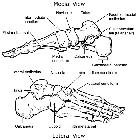
Tarsal bones
• Bones of the foot (Gray's illustration) - Ganzer Fall bei Radiopaedia
The metatarsals consist of five long bones in the foot. They are numbered from 1 to 5 from the medial side of the foot to lateral. They are analogous to the metacarpals of the hand.
Gross anatomy
The metatarsal bones run from the tarsus, forming the tarsometatarsal joints, to the base of proximal phalanges, forming the metatarsophalangeal joints.
Osteology
From proximal to distal, each metacarpal has a:
- base: articular surface to the tarsus
- body or shaft
- neck: expanded portion of the shaft approaching the head
- head: articular surface to the proximal phalanges
Articulations
- cuboid
- anterior surface articulates with the fourth and fifth metatarsal bones
- the facet for the fourth metatarsal is rectangular shaped
- the facet for the fifth metatarsal is triangular
- medial cuneiform
- articulates with the first and second metatarsals
- intermediate cuneiform
- articulates with the second metatarsal
- lateral cuneiform
- articulates with the second and third metatarsals
- phalanges
- the base of the proximal phalanges articulate with the hands of their corresponding metatarsal
Attachments
Musculotendinous
- tibialis anterior: inserts into a smooth facet at the anteroinferior angle of predominantly the medial cuneiform and also the adjacent first metatarsal
- peroneus longus: tendon winds around the tuberosity of the cuboid laterally to insert into the medial cuneiform and base of the first metatarsal, although predominantly the metatarsal
- peroneus tertius: inserts into the dorsum of the base of the fifth metatarsal and further forward anteriorly
- peroneus brevis: passes above peroneal trochlea to insert into base of the fifth metatarsal
- abductor digiti minimi: inserts into predominantly the proximal phalanx of the fifth toe, also onto the tubercle of the fifth metatarsal
- flexor hallucis longus: tendon runs along the bottom of the first metatarsal
- lumbricals: tendons lie on the plantar surface of the deep transverse ligament of the metatarsal heads
- adductor hallucis: oblique head arises anterior to flexor hallucis brevis, from the bases of the 2, 3 and 4 metatarsals
- flexor digiti minimi brevis: arises from the base of the fifth metatarsal bone and the adjoining fibrous sheath of peroneus longus, while the muscle belly runs along the fifth metatarsal
- plantar interossei: arise from the shaft of their own metatarsal and insert into the bases of the proximal phalanges
- dorsal interossei: arise by two heads from the two metatarsals between which it lies
- tibialis posterior: tendons insert into the navicular, but also given strong bands to the second, third and fourth metatarsals
Ligamentous
- transverse ligaments: bind the metatarsal heads together; the plantar aponeurosis slips insert into the edges of the transverse ligaments
- long plantar ligament: arises from the posterior tubercles of the calcaneus to insert into the bases of the central three metatarsal bones
Relations
Vascular
- dorsalis pedis: runs to base of the first intermetatarsal space
- lateral tarsal artery: runs laterally over bones of tarsus (just posterior to bases of metatarsals)
- arcuate artery: runs laterally beneath tendons of extensor digitorum brevis over bases of the metatarsal bones (further anteriorly compared to lateral tarsal artery)
- lateral plantar artery: crosses the sole, on the plantar foot, obliquely toward the base of the fifth metatarsal
- plantar arch: curves anteriorly forward across the bases of the fourth to second metatarsals
- dorsal metatarsal arteries: supply the 2, 3 and 4 intermetatarsal clefts
- dorsal venous arch: lies over heads of the metatarsals, draining into the medial and lateral ends of the great and small saphenous veins respectively
Nervous
- saphenous nerve: runs medially to reach as far as the head of the first metatarsal
- lateral plantar nerve: divides into superficial and deep branches at the fifth metatarsal
Muscular
flexor hallucis brevis: lies against the under surface of the first metatarsal, after arising from the under surface of the cuboid
Blood supply
- dorsal metatarsal arteries: arise from the arcuate artery and supply the 2-4 intermetatarsal clefts
- plantar metatarsal arteries: run forward from the plantar arch to supply the plantar side of the intermetatarsal clefts
- perforating arteries reinforce the supply from both
Structural considerations
The metatarsals significantly contribute to the arches of the foot:
- medial longitudinal arch: consists of calcaneus, talus, navicular, the three cuneiform bones and the 1 to 3 metatarsals
- lateral longitudinal arch: consists of calcaneus, cuboid and the 4th and 5th metatarsals
- transverse arch: consists of the bases of the five metatarsal bones and the adjacent cuboid and three cuneiforms
Development
Ossification
- metatarsals ossify by shaft centers in utero
- the epiphysis of the first metatarsal is at the base
- the epiphysis of the other four metatarsals are in the head

