Phalanges of the feet


The phalanges (single: phalanx) of the feet are the tubular bones of the toes. The second to fifth toes each contain a proximal, middle and distal phalanx whereas the great toe (hallux) only contains a proximal and distal phalanx.
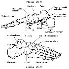
Gross anatomy
Osteology
Each phalanx consists of a central body or shaft and two extremities. The proximal phalanges have a long body that is convex superiorly and concave inferiorly. The two extremities are wider, with the base providing a concave articulation with the metatarsal heads and the head providing a convex articulation with the base of the middle phalanx (or distal phalanx in the case of the first toe). The middle phalanges are similarly shaped but much shorter. The distal phalanges are flat on the dorsal surface and taper towards the distal end, but then expanded at the distal extremity for support of the nail at the end of the toe .
Articulations
- metatarsophalangeal joint: between the metatarsals and proximal phalanges
- proximal interphalangeal joint: between the proximal and middle phalanges
- distal interphalangeal joint: between the middle and distal phalanges
- the great toe only has a single interphalangeal joint
Attachments
Musculotendinous
Extensor tendons attach on the dorsal aspect of the foot. In the great toe, the distal phalanx receives extensor hallucis longus whereas the proximal phalanx receives extensor hallucis brevis (part of extensor digitorum brevis). An extensor expansion also exists, formed from extensor digitorum, the lumbricals and interossei. The tendons of extensor digitorum brevis join the extensor expansion on the second, third and fourth toes .
On the plantar surface, flexor hallucis longus attaches to the base of the distal phalanx of the great toe and flexor hallucis brevis inserts, via the two sesamoids, into the sides of base of the proximal phalanx. The other four toes receive flexor digitorum brevis as it splits to insert onto the sides of the middle phalanx, and flexor digitorum longus passes through this to reach the distal phalanx. Abductor and flexor digiti minimi brevis also attach to the bases of the fifth proximal phalanx .
Ligamentous
Each MTP joint is strengthened by a thick pad of fibrocartilage (plantar ligament) on its plantar surface. Four short fibrous bands unite the plantar ligaments of adjacent MTP joints together to form the deep transverse metatarsal ligament. Collateral ligaments (similar to the hand) also reinforce the sides of the MTP and IP joints .
Blood supply
The lateral plantar artery gives rise to the single plantar arch of the foot. This curves convexly across the bases of the second, third and fourth metatarsals. From this arch, plantar metatarsal arteries run forwards and bifurcate to supply the four webs and digits. The medial plantar artery does not supply the plantar arch and instead supplies the great toe. Perforating arteries from the plantar arch and its metatarsal arteries reinforce the dorsal metatarsal arteries .
The veins accompanying the perforating arteries transport blood to the dorsal venous arch. The plantar muscles as a "sole pump" to aid returning venous blood flow .
Variant anatomy
Two sesamoid bones are often present present on the plantar aspect of the MTP joint of the great toe, of which the medial is the larger sesamoid (see multipartite hallux sesamoid). Sesamoid bones can also be found at the MTP joints of the second to fifth toes, and the IP joint of the great toe .
Development
Ossification
The phalanges are ossified from two centers :
- one for the shaft: appears at 10th week of gestation
- one for the base: appears between 4-10 years old, and fuses with the shaft around 18 years of age
Related investigations
Plain radiograph
The phalanges may be visualized on a number of series of the distal lower limb including:
Cross-sectional imaging
- CT
- MRI
Related pathology
- hallux valgus
- hallux rigidus
- trigger toe
- rheumatoid arthritis: musculoskeletal manifestations
- Rubinstein-Taybi syndrome
Siehe auch:

