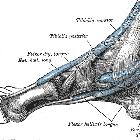
tibialis posterior


Tibialis
posterior muscle • Tibialis posterior tendon anatomy: diagram - Ganzer Fall bei Radiopaedia

Tibialis
posterior muscle • Lower leg axial section - Gray's anatomy illustration - Ganzer Fall bei Radiopaedia

Tibialis
posterior muscle • Plantar ligaments of the foot (Gray's illustration) - Ganzer Fall bei Radiopaedia

The tibialis posterior muscle is one of the small muscles of the deep posterior compartment of the leg.
Summary
- origin: upper half of posterior shaft of tibia and upper half of fibula between medial crest and interosseous border, and adjacent interosseous membrane.
- insertion: navicular and medial cuneiform
- the tendon splits into two slips after passing inferior to plantar calcaneonavicular ligament
- superficial slip inserts on the tuberosity of the navicular bone and sometimes medial cuneiform
- this is the main slip, accounting anterior two-thirds of the tendon
- deeper slip divides again into slips and has variable insertions onto the plantar surfaces of metatarsals 2 - 4, second cuneiform, cuboid, sustentaculum tali
- superficial slip inserts on the tuberosity of the navicular bone and sometimes medial cuneiform
- the tendon splits into two slips after passing inferior to plantar calcaneonavicular ligament
- action: plantarflexion and inversion of the foot
- arterial supply: posterior tibial artery
- innervation: tibial nerve
- antagonist: tibialis anterior
- variants:
- insertion into accessory navicular
Related pathology
Siehe auch:
und weiter:

 Assoziationen und Differentialdiagnosen zu Musculus tibialis posterior:


