Lateral cuneiform

Talus •
Lower limb bones (illustrations) - Ganzer Fall bei Radiopaedia

Lateral
cuneiform • Lateral cuneiform fracture - Ganzer Fall bei Radiopaedia

Tarsal bones
• Lateral cuneiform (Gray's illustration) - Ganzer Fall bei Radiopaedia
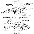
The lateral cuneiform is one of the tarsal bones located between the intermediate cuneiform and cuboid bones.
Gross anatomy
Osteology
The lateral cuneiform is a wedge-shaped bone. It is smaller than the medial cuneiform and larger than the intermediate cuneiform. It lies edge downward, between the intermediate cuneiform and cuboid.
Articulations
- anteriorly with the 2 and 3metatarsals
- posteriorly with the navicular
- laterally with the cuboid
- medially with the intermediate cuneiform
Attachments
Musculotendinous
- flexor hallucis brevis: the proximal part of the lateral cuneiform undersurface gives rise to this muscle
- tibialis posterior: one of its fibrous terminal tendon slips attaches to the narrow plantar surface
Ligamentous
- plantar interosseous ligaments: arise from the lower part of the lateral cuneiform, forming part of the transverse arch of the foot, these are mainly cuneocuboid ligament and intercuneiform ligaments which help gliding and rotation in pedal pronation or supination and also when the forefoot is stressed, as in initial thrust of running and jumping
Relations
- anterior: 2 and 3 metatarsal
- posterior: navicular
- lateral: cuboid
- medial: intermediate cuneiform
Blood supply
- via the lateral tarsal artery, a continuing branch of the dorsalis pedis
Development
Ossification
The lateral cuneiform ossifies in the first year of life.
Related pathology
Isolated lateral cuneiform fracture is rare, but has been documented in the literature ,
Siehe auch:
und weiter:

