hepatopulmonary syndrome


Hepatopulmonary syndrome (HPS) is a clinical syndrome defined by the presence of the following:
- liver disease
- dilation of pulmonary vasculature
- may involve pulmonary capillaries, pulmonary arteriovenous malformations, or pleural AVMs
- abnormalities in oxygenation
- elevation in the alveolar-arterial (A-a) oxygen gradient with or without hypoxemia on room air
Epidemiology
It is estimated to be present in ~15% of adults with liver cirrhosis.
Clinical presentation
Patients will classically present with varying degrees of positional dyspnea, alleivated by recubency and aggravated by assuming a seated position. Patients may bear the stigmata of concomitant liver disease, most commonly with a component of portal hypertension. The presence of severe hypoxemia with a measured partial pressure of arterial oxygen under 60 mmHg is particularly suggestive, as are spider nevi, clubbing of the digits, and cyanosis. Examination may also reveal:
- hepatosplenomegaly
- ascites, pedal edema
- caput medusae
- hyperdynamic circulation
- with an elevated cardiac output, decreased systemic vascular resistance, wide pulse pressure
- hypoxemia
- more profound when assuming a seated position
- with similarly positional dyspnea constitutes the platypnea-orthodeoxia syndrome
- may be absent due to compensation in mild disease, necessitating measurement of an A-a gradient
- more profound when assuming a seated position
Pathology
The characteristic vascular anomalies are attributed to a variety of mechanisms, including disregulation of pulmonary angiogenesis, an increase in pulmonary nitric oxide formation, and endothelial dysfunction.
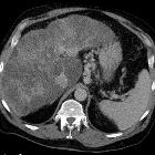
Radiographic features
The radiologic manifestations of this disease include distal vascular dilatation associated with an abnormally large number of visible terminal vessel branches, which are always concentrated in the lower lung zones.
Plain radiograph
Chest x-ray may include bibasal medium-sized (1.5-3 mm) nodular or reticulonodular opacities
CT
May show peripheral arteriolar dilatation on CT with an increased number of terminal branches extending towards the pleura.
Two patterns have been described on CTPA :
- type I
- most common (~85%)
- manifests as distal vascular dilatation with subpleural telangiectasia
- features include multiple, slightly dilated subpleural vessels that do not taper normally and therefore extend to the pleural surface
- type II
- less common (~15%)
- seen as individual arteriovenous malformations on angiograms and nodular dilatation of peripheral pulmonary vessels
Echocardiography
Contrast enhanced transthoracic echocardiography using agitated saline is a sensitive means of diagnosis, and will demonstrate passage of microbubbles into the left atrium within 3-6 cardiac cycles from opacification of the right atrium. The anatomic location of shunting will remain occult with this method . Transesophageal (TEE) studies may be more sensitive and allow direct visualization of microbubble influx in the left atrium from the pulmonary veins. The presence of esophageal varices, which have a high incidence in this population, warrant caution if TEE is to be performed .
Nuclear medicine - scintigraphy
Tc-m MAA (micro-aggregated albumin) lung scan is a useful method to detect intrapulmonary vascular dilatation. A scan showing uptake of radionuclide over the kidneys, brain, or both suggests shunting through the lung caused by an intrapulmonary shunt. The perfusion lung scan is a simple, safe, noninvasive and effective method to evaluate intrapulmonary vascular dilatation associated with right-to-left shunts.
See also
- pulmonary complications of cirrhosis and portal hypertension
- hepatopulmonary syndrome (HPS)
- portopulmonary hypertension (POPH)
- hepatic hydrothorax (HH)
Siehe auch:
und weiter:


 Assoziationen und Differentialdiagnosen zu Hepatopulmonales Syndrom: